. 2024 Oct 22;121(43):e2407838121.
doi: 10.1073/pnas.2407838121. Epub 2024 Oct 15.

A role for cross-linking proteins in actin filament network organization and force generation

Affiliations

A role for cross-linking proteins in actin filament network organization and force generation

Jennifer M Hill et al. Proc Natl Acad Sci U S A. .

Abstract

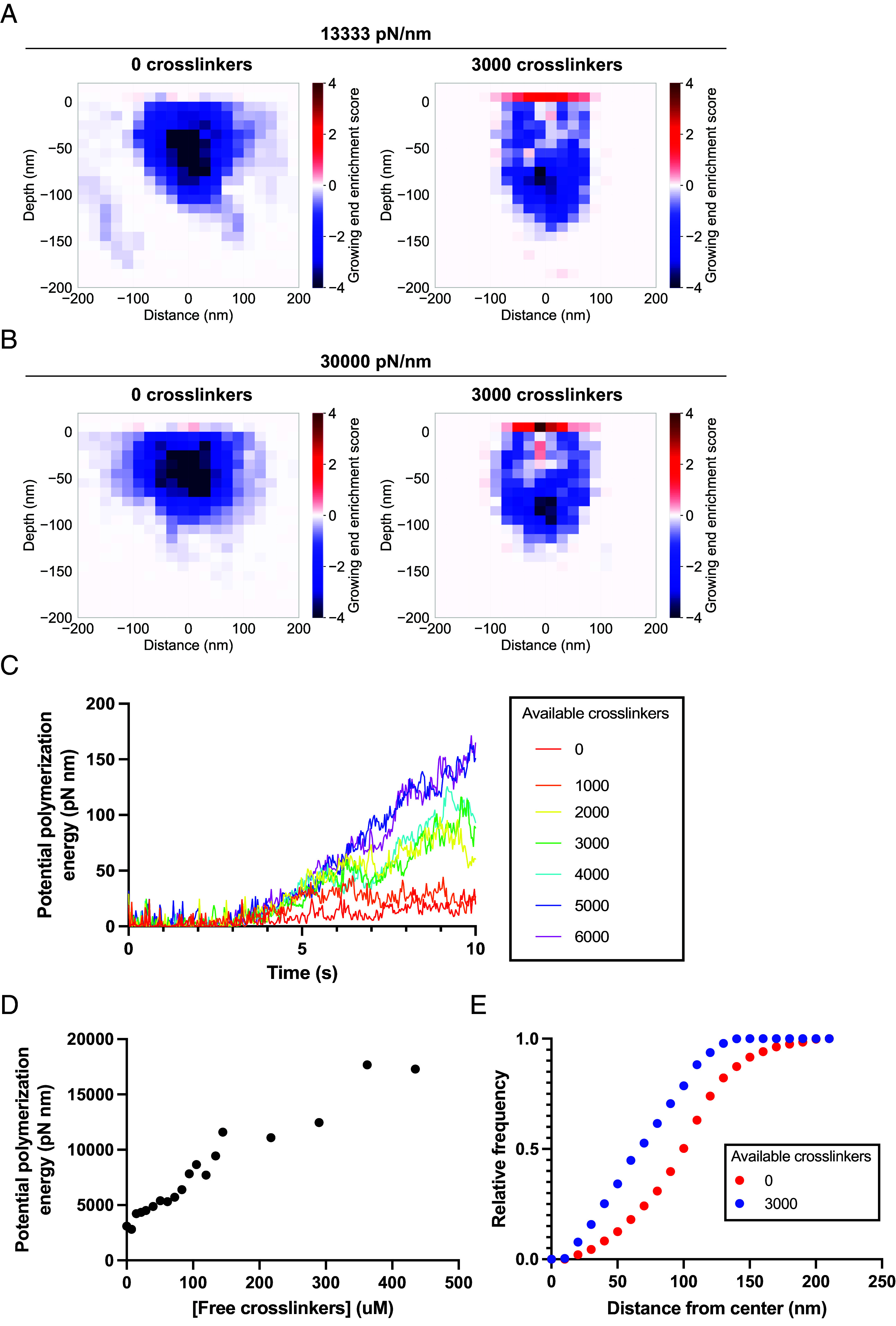
The high turgor pressure across the plasma membrane of yeasts creates a requirement for substantial force production by actin polymerization and myosin motor activity for clathrin-mediated endocytosis (CME). Endocytic internalization is severely impeded in the absence of fimbrin, an actin filament crosslinking protein called Sac6 in budding yeast. Here, we combine live-cell imaging and mathematical modeling to gain insights into the role of actin filament crosslinking proteins in force generation. Genetic manipulation showed that CME sites with more crosslinking proteins are more effective at internalization under high load. Simulations of an experimentally constrained, agent-based mathematical model recapitulate the result that endocytic networks with more double-bound fimbrin molecules internalize the plasma membrane against elevated turgor pressure more effectively. Networks with large numbers of crosslinks also have more growing actin filament barbed ends at the plasma membrane, where the addition of new actin monomers contributes to force generation and vesicle internalization. Our results provide a richer understanding of the crucial role played by actin filament crosslinking proteins during actin network force generation, highlighting the contribution of these proteins to the self-organization of the actin filament network and force generation under increased load.

Keywords: actin; clathrin-mediated endocytosis; crosslinking proteins; mathematical modeling.


Conflict of interest statement

Competing interests statement:The authors declare no competing interest.

Figures

Fig. 1.
Fig. 1.
Loss of fimbrin leads to an increase in the number of molecules of the minor actin filament crosslinking protein transgelin, but loss of transgelin does not affect the number of fimbrin molecules at CME sites. (A and B). Quantitative microscopy to determine the maximum number of GFP-tagged transgelin (A) or fimbrin (B) molecules at sites of CME in wild-type strains and strains lacking fimbrin (fimbrin) or transgelin (transgelin), respectively. Kymographs of individual sites show a slice from outside the cell (Top) to the inside (Bottom) over time and are not taken from the corresponding representative image. (C and D). Maximum number of molecules of transgelin (C) and fimbrin (D) at CME sites in wild-type and fimbrin or transgelin strains, respectively (n = 90 sites from ≥10 cells per strain). (E). Representative images of mCherry-tagged Sla1 and GFP-tagged transgelin in WT, fimbrin, and fimbrin transgelin overexpression (transgelin OE) strains, 24 h postinduction with β-estradiol. Sites where Sla1 and transgelin hook into the cell in kymographs were considered internalized. Representative kymographs are shown for all strains and are not taken from the corresponding representative image. (F). Quantification of maximum numbers of transgelin molecules arriving at CME sites in WT, fimbrin, and fimbrin transgelin OE strains, 24 h postinduction with β-estradiol (n = 60 sites from ≥10 cells per strain). (G). Fraction of CME sites internalized in WT, fimbrin, and fimbrin transgelin OE strains, 24 h postinduction with β-estradiol (n = 60 sites from ≥10 cells per strain). Error bars show SD.
Fig. 2.
Fig. 2.
Mathematical modeling reveals that crosslinking proteins contribute to actin network force generation for membrane internalization during CME. (A) Schematic of an agent-based model for the actin network associated with CME sites (9). The endocytic vesicle is modeled as a bead on a spring (blue) that resists internalization. Actin filaments attached to the vesicle generate force for internalization by polymerizing at the membrane. (B) Final frame of simulations with pools of either 0 or 3,000 available crosslinking proteins. The view shows a slice from the center of the network to reveal the internalized vesicle in the network (blue). Cyan pointers indicate filaments that have dissociated from the network. (C) Numbers of total and double-bound crosslinkers in the final actin network across a range of free crosslinker concentrations. N = 5 simulations, error bars show SD. (D) Scatter plot of the 90th percentile of the extent of vesicle internalization for simulations with a range of numbers of double-bound crosslinkers in the final network. The dotted line at Y = 60 nm represents threshold for snap-through transition and scission. (E) Scatter plot of the 90th percentile of the extent of vesicle internalization for simulations with varying numbers of total crosslinkers in the final network. The dotted line at Y = 60 nm represents threshold for snap-through transition and scission. (F) Scatter plot of the 90th percentile of the extent of internalization for individual simulations with varying numbers of double-bound crosslinkers and membrane resistances. Solid lines show linear regressions for each set of simulations at a given membrane resistance.
Fig. 3.
Fig. 3.
The extent to which CME site internalization is hindered by hypotonic shock depends on crosslinking protein abundance. Cells were grown in media containing 1 M sorbitol and shifted to media containing 0.25 M sorbitol for 5 min. All strains are in a background containing a deletion of the genes encoding transgelin and Fps1. SAC6 dupl.: duplication of the gene encoding fimbrin at the endogenous locus. (A) Quantitative imaging of GFP-tagged fimbrin 5 min after osmotic shock. (B) Imaging of GFP-tagged Sla1 and mRFP-tagged Abp1 5 min after osmotic shock. Sites where Sla1 and Abp1 hook into the cell in kymographs were considered internalized. Representative kymographs are shown for all strains and are not taken from the corresponding representative image. (C) Fraction of endocytic sites that are internalized as a function of the maximum number of fimbrin molecules at endocytic sites 5 min postshock (n = 90 sites from ≥10 cells per strain). Error bars show SD.
Fig. 4.
Fig. 4.
Endocytic sites that internalize under hypotonic shock conditions have a higher maximum number of fimbrin molecules than sites that stall. (A) Cells were grown in 1 M sorbitol media and shifted to 0.25 M sorbitol media. Fimbrin and Abp1 were imaged after 5 min. Representative kymographs are shown for sites that internalize and fail and are not taken from the corresponding representative image. (B) Maximum number of fimbrin molecules at endocytic sites that internalize (n = 62 sites) or fail (n = 38 sites). (C) Maximum fluorescence intensity of mRFP-tagged Abp1 at endocytic sites that internalize (n = 62 sites) or fail (n = 38 sites). (D) Ratio of the maximum number of fimbrin molecules to Abp1-mRFP fluorescence intensity at endocytic sites that internalize (n = 62 sites) or fail (n = 38 sites). Error bars show SD.
Fig. 5.
Fig. 5.
Mathematical modeling predicts self-organization of actin filaments to maximize force production. (A and B) Heat map of enrichment scores for growing ends relative to capped ends of actin filaments in the final network with pools of either 0 or 3,000 available crosslinking proteins and 13,333 pN/nm (A) or 30,000 pN/nm (B) required to internalize the vesicle. N = 5 simulations. (C) Average potential polymerization energy of the actin filament network over the duration of simulations with varying pools of available crosslinking proteins. N = 5 simulations. (D) Total potential polymerization energy of the actin filament network as a function of the concentration of free crosslinkers. N = 5 simulations. (E) Cumulative distribution functions for the relative frequency of plus ends measured radially from the center of the vesicle for simulations with pools of 0 and 3,000 available crosslinking proteins. N = 5 simulations.

Update of

References

    1. Kaksonen M., Toret C. P., Drubin D. G., A modular design for the clathrin- and actin-mediated endocytosis machinery. Cell 123, 305–320 (2005). - PubMed
    1. Goode B. L., Eskin J. A., Wendland B., Actin and endocytosis in budding yeast. Genetics 199, 315–358 (2015). - PMC - PubMed
    1. Boettner D. R., Chi R. J., Lemmon S. K., Lessons from yeast for clathrin-mediated endocytosis. Nat. Cell Biol. 14, 2–10 (2011). - PMC - PubMed
    1. Merrifield C. J., Feldman M. E., Wan L., Almers W., Imaging actin and dynamin recruitment during invagination of single clathrin-coated pits. Nat. Cell Biol. 4, 691–698 (2002). - PubMed
    1. Kaksonen M., Sun Y., Drubin D. G., A pathway for association of receptors, adaptors, and actin during endocytic internalization. Cell 115, 475–487 (2003). - PubMed

MeSH terms

LinkOut - more resources