. 2024 Oct 29;18(43):29439-29456.
doi: 10.1021/acsnano.3c12337. Epub 2024 Oct 15.

Robust and Sustained STING Pathway Activation via Hydrogel-Based In Situ Vaccination for Cancer Immunotherapy

Affiliations

Robust and Sustained STING Pathway Activation via Hydrogel-Based In Situ Vaccination for Cancer Immunotherapy

Sheng-Liang Cheng et al. ACS Nano. .

Abstract

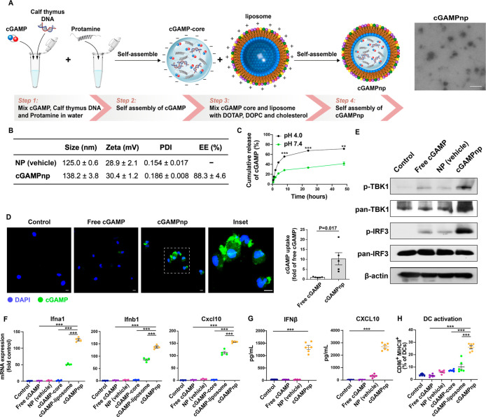
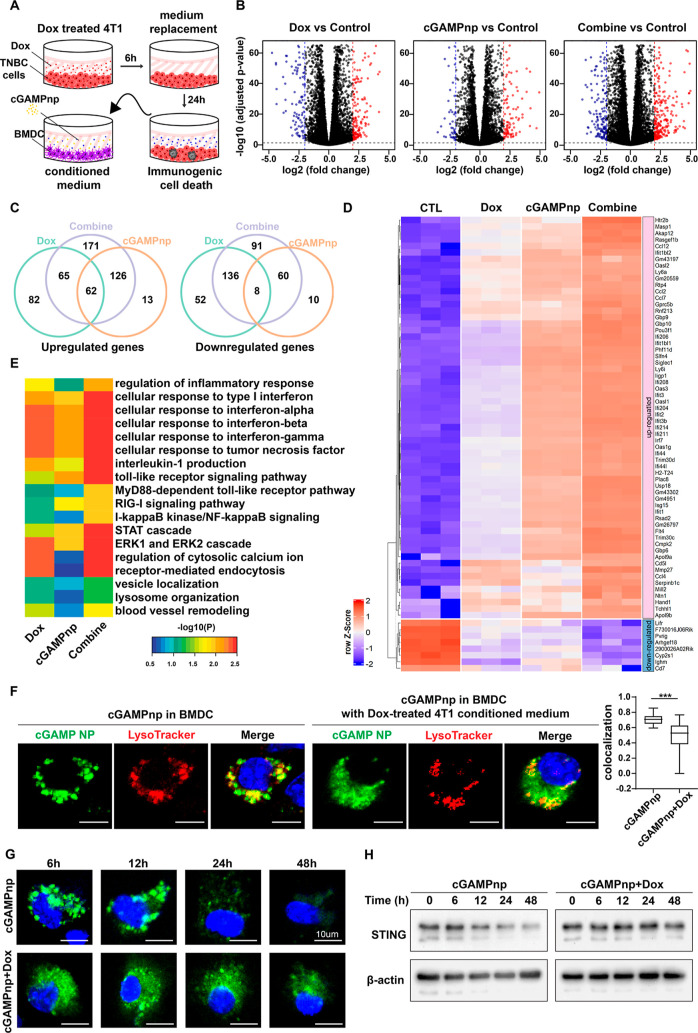
The stimulator of interferon genes (STING) pathway is crucial for tumor immunity, leading to the exploration of STING agonists as potential immunotherapy adjuvants. However, their clinical application faces obstacles including poor pharmacokinetics, transient activation, and an immunosuppressive tumor microenvironment (TME). Addressing these limitations, our study aims to develop an injectable silk fibroin hydrogel-based in situ vaccine. It incorporates a nanoscale STING agonist, an immunogenic cell death (ICD) inducer, and an immunomodulator to ensure their controlled and sustained release. cGAMP nanoparticles (cGAMPnps) with a core-shell structure ensure optimal delivery of cGAMP to dendritic cells (DCs), thereby activating the STING pathway and fostering DC maturation. ICD-associated damage-associated molecular patterns amplify and prolong STING activation via enhanced type I IFN and other inflammatory pathways, along with delayed degradation of cGAMP and STING. Furthermore, the STING-driven vascular normalization by cGAMPnps and ICD, in conjunction with immunomodulators like antiprogrammed cell death protein 1 antibody (anti-PD-1 Ab) or OX40 ligand (OX40L), effectively remodels the immunosuppressive TME. This in situ gel vaccine, when used independently or with surgery as neoadjuvant/adjuvant immunotherapy, enhances DC and CD8+ T-cell activation, suppressing tumor progression and recurrence across various immunologically cold tumor models. It revolutionizes the application of STING agonists in cancer immunotherapy, offering substantial promise for improving outcomes across a broad spectrum of malignancies.

Keywords: STING agonist; cGAMP; cancer immunotherapy; immunogenic cell death; nanoparticle; neoadjuvant immunotherapy; silk fibroin hydrogel.


Conflict of interest statement

The authors declare no competing financial interest.

Figures

Figure 1
Figure 1
Schematic representation of in situ gel vaccination for the sustained release of cGAMPnps, Dox, and an immunomodulator to generate durable STING activation and effective anticancer immunity. (A) Thermoresponsive SF hydrogel-based in situ gel vaccine encapsulates a nanoscale STING agonist (cGAMPnp), an ICD inducer, and an immunomodulator, providing a localized depot for the controlled, sustained release of immunotherapeutic agents. The in situ gel vaccine effectively triggers STING-driven tumor vascular normalization and activates DCs and effector CD8+ T cells within tumors, as well as in their draining lymph nodes and distal metastases. This results in the suppression of tumor progression and prevention of recurrence in immunologically cold tumors. (B) Nanoscale STING agonist cGAMPnps promotes the efficient uptake of cGAMP and induces activation of the type I IFN response in DCs. The hydrogel formulation containing ICD inducer Dox triggers release of tumor antigens and enhances and prolongs STING activation by cGAMPnps. Furthermore, checkpoint inhibitors (e.g., anti-PD-1 antibody) or immunomodulators (OX40L) incorporated into the hydrogel reprogram the immunosuppressive TME, enhancing the efficacy of in situ cancer vaccination.
Figure 2
Figure 2
cGAMP-loaded NPs (cGAMPnps) enhance intracellular cGAMP uptake and STING activation in DCs. (A) Schematic illustration of the synthesis procedure and representative TEM image of cGAMPnps. Scale bar, 200 nm. (B) Size, zeta potential, polydispersity index (PDI), and EE of the cGAMPnp or empty NP (vehicle) (n = 3). (C) Kinetics of cGAMP release from NPs under physiological (pH 7.4) or acidic (pH 4.0) conditions. The release of cGAMP was measured as the fluorescence intensity of fluorescein-labeled cGAMP (n = 3). (D) Fluorescein-cGAMP uptake by bone marrow-derived dendritic cells (BMDCs) treated with cGAMPnps or free cGAMP. The cellular uptake of fluorescein-cGAMP (1 μM) was imaged and quantified using a Zeiss LSM 780 confocal microscope (n = 5). (E) Western blotting was utilized to analyze TANK-binding kinase 1 (TBK1) and Interferon Regulatory Factor 3 (IRF3) phosphorylation in BMDCs treated with free cGAMP, empty vehicle, or cGAMPnps (1 μM). (F) mRNA expression levels of type I IFN and inflammation-related genes (Ifna1, Ifnb1, and Cxcl10) in BMDCs 6 h after treatment with free cGAMP, empty vehicle, cGAMP-DNA-protamine core (cGAMP-core), cGAMP loaded in the liposome (cGAMP-liposome), or cGAMPnps (0.5 μM) were measured by RT–qPCR. The results are expressed as the fold change relative to the corresponding level in the untreated control group (n = 4). (G) Cytokine secretion from BMDCs was measured by ELISA 24 h after treatment with free cGAMP, empty vehicle or cGAMPnps (0.5 μM) (n = 6). (H) Expression of MHC-II and CD86 in CD11c+ BMDCs was measured by flow cytometry 24 h after treatment with free cGAMP, empty vehicle, cGAMP-core, cGAMP-liposome, or cGAMPnps (1 μM) (n = 6). All data are shown as the mean ± standard error of the mean (SEM) *P < 0.05, **P < 0.01, ***P < 0.001.
Figure 3
Figure 3
Gene expression changes and ontological analysis in BMDCs treated with cGAMPnps and ICD-associated DAMPs. (A) Schematic representation of the experimental design. 4T1 cells were treated with Dox (2 μM) for 6 h, the medium was replaced with fresh medium, and the conditioned medium was collected after 24 h. BMDCs were treated with cGAMPnps (1 μM) alone or with the collected the conditioned medium. (B) Heatmap showing the differentially expressed genes in BMDCs upon treatment with cGAMPnps, ICD-associated DAMPs from Dox-treated 4T1 cells (Dox), or the combination of both compared to the untreated control group (n = 3). (C) Venn diagram showing the number of differentially expressed genes among different treatment groups. (D) Heatmap showing the expression profiles of common genes among different treatment groups (n = 3). (E) Gene ontological analysis of differentially expressed genes related to inflammatory and STING pathway activation (n = 3). (F) Confocal microscopy images of BMDCs treated with cGAMPnps (green) alone or in combination with ICD-associated DAMPs and stained with Lysotracker Red DND-99 (red). BMDCs were treated with or without the conditioned medium from 4T1 breast cancer cells exposed to Dox for 24 h and then incubated with cGAMPnps loaded with fluorescein-cGAMP for 6 h; subsequently, the uptake and trafficking of cGAMP in BMDCs was examined. Scale bar, 10 μm. The colocalization of fluorescein-cGAMP with Lysotracker Red DND-99 in BMDCs was quantified (n = 15). Data are shown as the mean ± SEM ***P < 0.001. (G) BMDCs treated by cGAMPnps with or without the conditioned medium from 4T1 breast cancer cells exposed to Dox exhibit different temporal patterns of cGAMP-punctate formation and depletion. Scale bar, 10 μm. (H) Western blot analysis of STING degradation in BMDCs treated with cGAMPnp (1 μM) alone or in combination with ICD-associated DAMPs from Dox-treated 4T1 cells (Dox) for different time points. All data are shown as the mean ± SEM.
Figure 4
Figure 4
Enhanced and prolonged STING activation in BMDCs treated with cGAMPnps and ICD-associated DAMPs. (A) Western blot analysis of TBK1 and IRF3 phosphorylation in BMDCs treated with cGAMPnp (1 μM), ICD-associated DAMPs from Dox-treated 4T1 cells (Dox), or the combination of both for 6 or 24 h, as described in Figure 3a. (B) Western blot analysis of TBK1 and IRF3 phosphorylation in BMDCs treated with cGAMPnp (1 μM) alone or in combination with ICD-associated DAMPs from Dox-treated 4T1 cells (Dox) for different time points. (C) mRNA expressions of type I IFN and inflammation-related genes (Ifna1, Ifnb1, Tnf, and Cxcl10) in BMDCs 6, 24, or 48 h after treatment with cGAMPnp (1 μM), ICD-associated DAMPs from Dox-treated 4T1 cells (Dox), or the combination of both were measured by RT–qPCR. The results are expressed as the fold change relative to the corresponding level in the untreated control group (n = 6). (D) ELISA analysis of IFN-β and CXCL10 production in BMDCs treated with cGAMPnp (1 μM), ICD-associated DAMPs from Dox-treated 4T1 cells (Dox), or the combination of both for 6 or 24 h (n = 5). (E) Flow cytometry analysis of MHC II and CD86 expression in BMDC cells treated with cGAMPnp (1 μM), ICD-associated DAMPs from Dox-treated 4T1 cells (Dox), or the combination of both for 24 h (n = 6). ns, not significant. All data are shown as the mean ± SEM *P < 0.05, **P < 0.01, ***P < 0.001, ****P < 0.0001.
Figure 5
Figure 5
Injectable SF hydrogels enable a sustained release of cGAMPnps and Dox to potentiate STING activation in TNBC tumors. (A) Photographs of SF in various stages of the gelation process. (B) SEM images of the SF hydrogel before and after loading with cGAMPnps and Dox. (C) Release of fluorescein-cGAMP loaded cGAMPnp and Dox from SF hydrogels under physiological conditions (pH 7.4) and acidic conditions (pH 6.8) (n = 3). (D) In vivo fluorescent imaging of 4T1 tumor-bearing mice at 4, 8, 24, and 48 h after intratumoral injection of free Dox, free fluorescein-cGAMP or Dox, or fluorescein-cGAMPnp loaded SF hydrogels. (E) Quantification analysis of the retention of Dox and fluorescein-cGAMP in tumor tissues after injection of free drugs or hydrogels (n = 3). (F) Representative confocal images of the intracellular uptake of cGAMP by tumors. Green, cGAMP; blue, DAPI. Scale bar, 50 μm. (G) Flow cytometry was used to analyze the immune cell types that take up fluorescein-cGAMP in the 4T1 tumors 2 h after injection of free fluorescein-cGAMP or fluorescein-cGAMPnp-loaded SF hydrogels. DCs, macrophages, and monocytes were identified as CD45+CD11c+, CD45+F4/80+, and CD45+CD11b+ cells, respectively, and other lymphocytes were identified as CD3+CD4+ cells and CD3+CD8+ cells. (H) mRNA expressions of type I IFN and inflammation-related genes (Ifna1, Ifnb1, Tnf, and Cxcl10) in 4T1 tumors and TDLNs 24 h after intratumoral injection of Dox@Gel, cGAMPnp@Gel, or Dox-cGAMPnp@Gel (n = 7). All data are shown as the mean ± SEM *P < 0.05, **P < 0.01, ***P < 0.001.
Figure 6
Figure 6
Effect of the in situ gel vaccine (Dox-cGAMPnp@Gel) on vessel normalization and activation of anticancer immunity in an orthotopic 4T1 TNBC model. (A) Schematic of the experimental protocol. Various formulations were intratumoral administered on days 9, 16, and 23 after the orthotopic inoculation of 4T1 cells, and tumors and TDLNs were analyzed on day 30. (B) Pericyte coverage in TNBC was imaged and quantified using a Zeiss LSM 780 confocal microscope (n = 4). CD31-positive endothelial cells are stained green, and NG2-positive pericytes are stained red. Scale bar, 40 μm. (C) Expression of the antiangiogenic and vessel stabilizing factors in BMDCs after treatment with cGAMPnps with the conditioned medium from Dox-treated 4T1 cells measured through RNA-seq (n = 3). (D) Hoechst 33342-positive cells (n = 8), DCs (CD45+CD11c+), activated DCs (CD86+MHCII+), and cytotoxic CD8+ T lymphocytes (CD3+CD8+) in tumors were detected by flow cytometry (n = 10). (E) DCs (CD45+CD11c+), activated DCs (CD86+MHCII+), and cytotoxic CD8+ T lymphocytes (CD3+CD8+) in TDLNs were detected by flow cytometry (n = 10). (F) Percentages of central memory (CD44+CD62L+) CD8 T cells in spleens were detected by flow cytometry (n = 8). (G) Tumor growth curve of the 4T1 TNBC model after treatment with various formulations (n = 8). (H) Number of spontaneously occurring lung metastatic nodules in the orthotopic TNBC (4T1) model was reduced in mice treated with cGAMPnp@Gel or Dox-cGAMPnp@Gel (n = 21). Images of hematoxylin and eosin (H&E) staining show metastatic tumor nodules in the lung. Scale bar, 200 μm. (I) Schematic of the experimental protocol. Nine days after the orthotopic inoculation of 4T1 cells, mice were intratumorally administered with various formulations. The implanted tumor was surgically removed and various formulations were injected into the surgical site on day 16. (J) Tumor growth curve and overall survival in the postsurgical 4T1 TNBC model after treatment with various formulations (n = 12). The blue arrow shows surgical removal of primary tumors and the red arrow shows administration of various formulations. A comparison of survival curves was performed using a log-rank Mantel–Cox test (two-sided). All data are shown as the mean ± SEM *P < 0.05, **P < 0.01, ***P < 0.001.
Figure 7
Figure 7
In situ gel vaccine containing cGAMPnps, Dox, and an immunomodulator (anti-PD-1 antibody or OX40L) achieves potent antitumor immunity and prevents postsurgical tumor recurrence in orthotopic TNBC and GBM models. (A) Release of FITC-BSA from SF hydrogels under physiological conditions (pH 7.4) and acidic conditions (pH 6.8) (n = 3). (B) In vivo fluorescent imaging of 4T1 tumor-bearing mice at 4, 8, 24, and 48 h after intratumoral injection of free FITC-BSA, or FITC-BSA-loaded SF hydrogels. Quantification analysis of the retention of FITC-BSA in tumor tissues (n = 3). (C) Schematic of the experimental protocol. For the orthotopic TNBC model, 9 days after the orthotopic inoculation of 4T1 cells, mice were intratumorally administered with various formulations. The implanted tumor was surgically removed, and various formulations were injected into the surgical site on day 16, and tumors and TDLNs were analyzed on day 23. For the orthotopic GBM model, 12 days after the orthotopic inoculation of ALTS1C1 cells, the implanted tumor was surgically removed and various formulations were injected into the surgical site. Tumors were analyzed on day 26. (D) DCs (CD45+CD11c+), activated DCs (CD86+MHCII+), cytotoxic CD8+ T lymphocytes (CD3+CD8+), and activated CD8+ T lymphocytes (IFN-γ+ in CD8+) in TDLNs of 4T1 tumor-bearing mice were detected by flow cytometry (n = 8). (E) Cytotoxic CD8+ T lymphocytes (CD3+CD8+) and activated CD8+ T lymphocytes (IFN-γ+ in CD8+) in lungs of 4T1 tumor bearing mice were detected by flow cytometry (n = 8). (F) Growth curve shows the primary tumor regrowth postresection in the 4T1 TNBC model. (G) Overall survival in the 4T1 TNBC model (n = 12). (H) DCs (CD45+CD11c+), activated DCs (CD86+MHCII+), cytotoxic CD8+ T lymphocytes (CD3+CD8+), and activated CD8+ T lymphocytes (IFN-γ+ in CD8+) in ALTS1C1 GBM were detected by flow cytometry (n = 5). (I) Tumor volume in the GBM model in different treatment groups (n = 7). (J) Overall survival in the GBM model (n = 10). The blue arrow shows surgical removal of primary tumors, and the red arrow shows administration of various formulations. A comparison of survival curves was performed using a log-rank Mantel–Cox test (two-sided). All data are shown as the mean ± SEM *P < 0.05, **P < 0.01, ***P < 0.001.
Figure 8
Figure 8
In situ gel vaccine containing cGAMPnp, Dox, and anti-PD-1 antibody achieves potent antitumor immunity in orthotopic HCC and PDAC models. (A) Schematic of the experimental protocol. Five days after the orthotopic implantation of murine HCC (HCA-1) cells, mice were treated with various formulations on days 5, 12, and 19, and tumor size was measured on day 24. Four days after the orthotopic implantation of murine PDAC (KPC001) cells, mice were treated with various formulations on days 4, 8, and 12, and tumor size was measured on day 16. cGAMP (0.8 mg/kg), Dox (2 mg/kg), and anti-PD-1 antibody (6 mg/kg) loaded in SF gels were intraperitoneally injected into orthotopic HCC (HCA-1) and PDAC (KPC001) models. (B–E) DCs (CD45+CD11c+) (B), activated DCs (CD86+MHCII+) (C), cytotoxic CD8+ T lymphocytes (CD3+CD8+) (D), and activated CD8+ T lymphocytes (IFN-γ+ in CD8+) (E) in HCC and PDAC were detected by flow cytometry (n = 8). (F) Tumor volume in the orthotopic HCC and PDAC models in different treatment groups (n = 10). (G) Representative immunofluorescence images and quantification of TUNEL staining (green) with DAPI counterstain (blue) in tumors after treatment of the orthotopic HCC and PDAC models with different treatments, as described in a (n = 7 images from four mice). Scale bar, 10 μm. (H) Number of spontaneously occurring lung metastatic nodules in the orthotopic HCC (HCA-1) model was reduced in mice treated with Dox-cGAMPnp@Gel or aPD1-Dox-cGAMPnp@Gel (n = 24). Images of H&E staining of metastatic tumor nodules in the lung. Scale bar, 200 μm. (I) Experimental design to examine the translational potential of the in situ gel vaccine by evaluating STING activation in human tissues. (J) mRNA expression of type I IFN-related genes (IFNA1 and IFNB1) in PDAC patient tumor samples after treatment with cGAMPnp (1 μM) with or without combination of Dox (2 μM) for 6 h. All data are shown as the mean ± SEM *P < 0.05, **P < 0.01, ***P < 0.001, ****P < 0.0001.

References

    1. Yi Y.; Yu M.; Li W.; Zhu D.; Mei L.; Ou M. Vaccine-like nanomedicine for cancer immunotherapy. J. Controlled Release 2023, 355, 760–778. 10.1016/j.jconrel.2023.02.015. - DOI - PubMed
    1. Jiang Z.; Xu Y.; Du G.; Sun X. Emerging advances in delivery systems for mRNA cancer vaccines. J. Controlled Release 2024, 370, 287–301. 10.1016/j.jconrel.2024.04.039. - DOI - PubMed
    1. Saxena M.; van der Burg S. H.; Melief C. J. M.; Bhardwaj N. Therapeutic cancer vaccines. Nat. Rev. Cancer 2021, 21 (6), 360–378. 10.1038/s41568-021-00346-0. - DOI - PubMed
    1. Lin M. J.; Svensson-Arvelund J.; Lubitz G. S.; Marabelle A.; Melero I.; Brown B. D.; Brody J. D. Cancer vaccines: the next immunotherapy frontier. Nat. Cancer 2022, 3 (8), 911–926. 10.1038/s43018-022-00418-6. - DOI - PubMed
    1. Viswanath D. I.; Liu H. C.; Huston D. P.; Chua C. Y. X.; Grattoni A. Emerging biomaterial-based strategies for personalized therapeutic in situ cancer vaccines. Biomaterials 2022, 280, 121297.10.1016/j.biomaterials.2021.121297. - DOI - PMC - PubMed

Publication types

MeSH terms

LinkOut - more resources