. 2025 Apr;72(4):393-407.
doi: 10.1165/rcmb.2024-0153OC.

MrgprC11+ Jugular Neurons Control Airway Hyperresponsiveness in Allergic Airway Inflammation

Affiliations

MrgprC11+ Jugular Neurons Control Airway Hyperresponsiveness in Allergic Airway Inflammation

Yanyan Xing et al. Am J Respir Cell Mol Biol. 2025 Apr.

Abstract

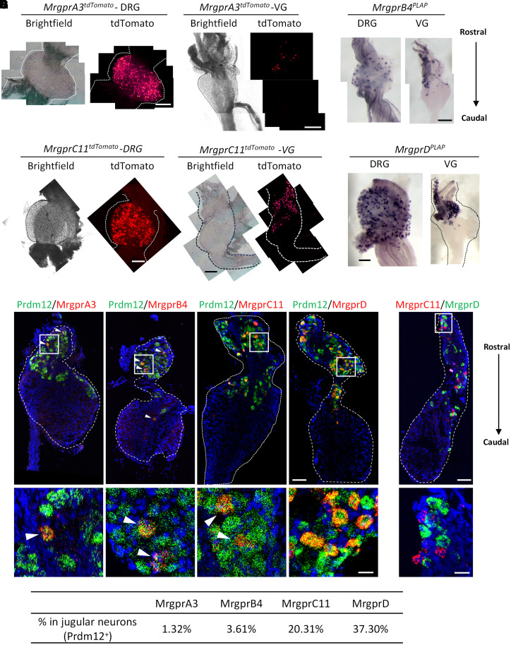
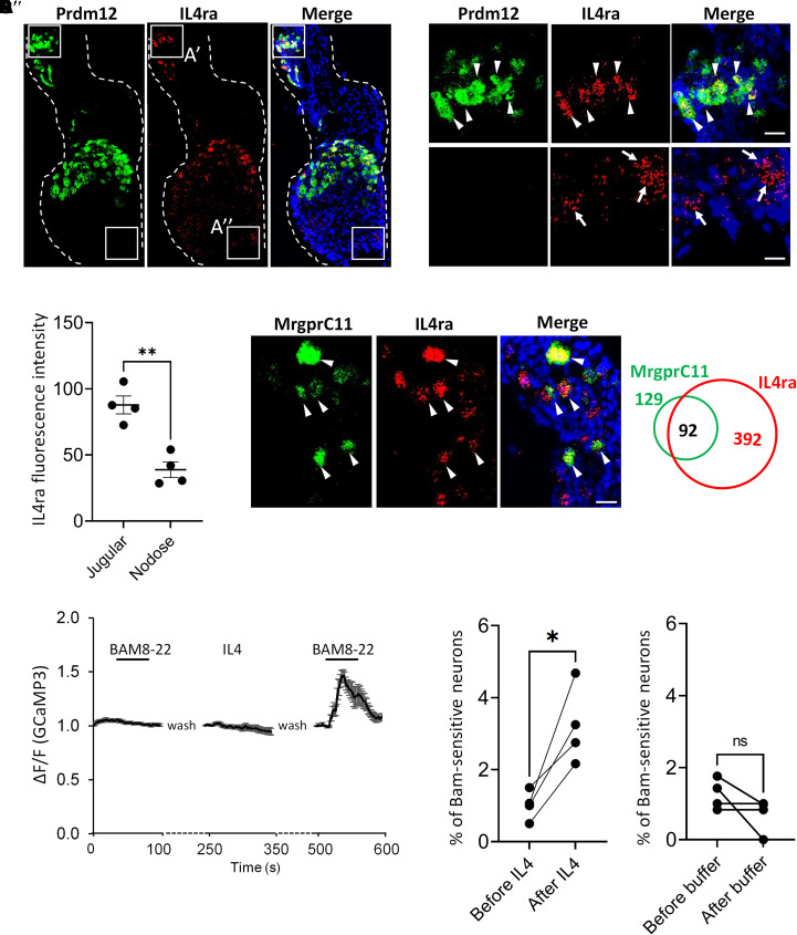
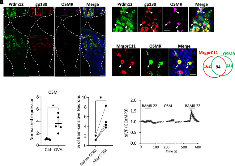
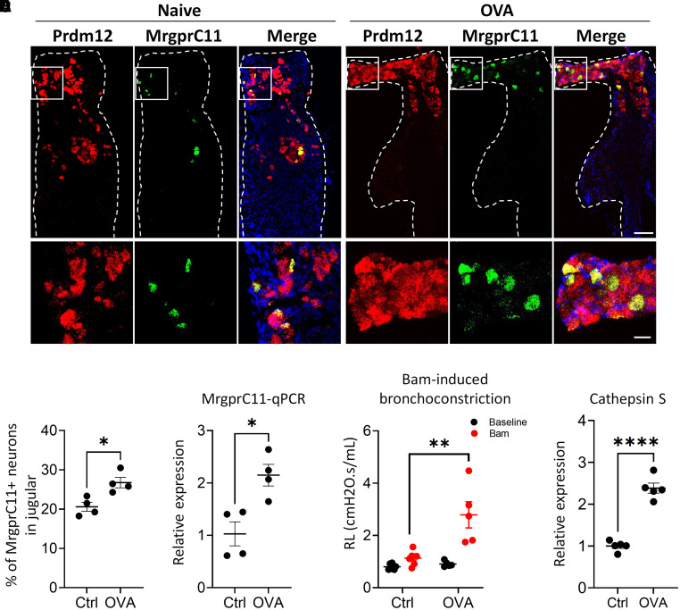
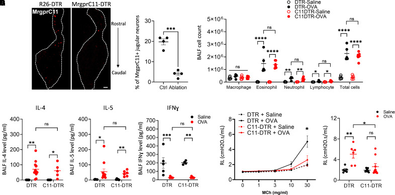
The lung is densely innervated by sensory nerves, the majority of which are derived from the vagal sensory neurons. Vagal ganglia consist of two different ganglia, termed nodose and jugular ganglia, with distinct embryonic origins, innervation patterns, and physiological functions in the periphery. Because nodose neurons constitute the majority of the vagal ganglia, our understanding of the function of jugular nerves in the lung is very limited. This study aims to investigate the role of MrgprC11+ jugular sensory neurons in a mouse allergic asthma model. Our previous study has shown that MrgprC11+ jugular neurons mediate cholinergic bronchoconstriction. In this study, we found that, in addition to MrgprC11, several other Mrgpr family members, including MrgprA3, MrgprB4, and MrgprD, are also specifically expressed in the jugular sensory neurons. MrgprC11+ jugular neurons exhibit dense innervation in the respiratory tract, including the larynx, trachea, proximal bronchus, and distal bronchus. We also found that receptors for IL-4 and oncostatin M, two critical cytokines promoting allergic airway inflammation, are mainly expressed in jugular sensory neurons. Both IL-4 and oncostatin M can sensitize the neuronal responses of MrgprC11+ jugular neurons. Moreover, ablation of MrgprC11+ neurons significantly inhibited airway hyperresponsiveness in the asthmatic lung, demonstrating the critical role of MrgprC11+ neurons in controlling airway constriction. Our results emphasize the critical role of jugular sensory neurons in respiratory diseases and present MrgprC11+ neurons as a potential therapeutic target for treating airway hyperresponsiveness.

Keywords: airway hyperresponsiveness; allergic asthma; jugular neurons; neuroimmune interactions; vagal sensory neurons.


Figures

Figure 1.
Figure 1.
Mrgprs are specifically expressed in jugular sensory neurons. (A–D) Expression of MrgprA3 (A), MrgrpB4 (B), MrgprC11 (C), and MrgprD (D) in both dorsal root ganglia (DRG) and vagal ganglia (VG) using mouse genetic labeling. Mrgprs+ DRG sensory neurons are evenly distributed within the ganglia. However, all Mrgprs+ vagal sensory neurons are located in the rostral part of the ganglia. Brightfield images were collected for tdTomato labeling to visualize the morphology of the ganglia. (E) RNAscope in situ analysis showing that Mrgprs are only expressed in jugular sensory neurons labeled by Prdm12. Arrowheads point to some of the overlapping cells. DAPI–nuclei stain is indicated in blue. (F) The expressions of MrgprC11 and MrgprD define two different jugular nociceptive populations. The boxed areas in the upper panels in (E) and (F) are shown at greater magnification in the lower panels. (G) Table showing the percentage of jugular neurons expressing Mrgpr family members. Scale bars: A–D, 250 μm; E and F, 100 μm (upper panels); E and F, 25 μm (lower panels).
Figure 2.
Figure 2.
Whole-mount placental alkaline phosphatase (PLAP) histochemistry analysis of the airway isolated from MrgprC11PLAP mice with tamoxifen treatment to examine the innervation of MrgprC11+ nerves. (A–C) MrgprC11PLAP mice were treated with a high dose of tamoxifen (100 mg/kg, once daily for 5 days) to visualize all MrgprC11+ nerves. Dense nerves were observed in the (A) larynx, (B) trachea, and (C) main bronchi. The trachea was cut along the anterior midline of the cartilage to make a flat whole mount. (D and E) MrgprC11PLAP mice were treated with a low dose of tamoxifen (10 mg/kg, once) to sparsely label a subset of MrgprC11+ neurons. Terminal arbors were observed in both the (D) proximal large airway and (E) distal small airway. (F) Lung sections collected from MrgprC11tdTomato mice that were treated with a high dose of tamoxifen. MrgprC11+ lung interoceptors penetrate into the superficial luminal surface. DAPI–nuclei stain is indicated in blue. Scale bars: A–C, 1 mm; D and E, 0.1 mm; F, 50 μm.
Figure 3.
Figure 3.
MrgprC11+ jugular neurons innervate the lung. (A) Diagram illustrating injection of adeno-associated virus–enhanced GFP (AAV-EGFP) and AAV-flex-tdTomato into the VG of MrgprC11CreER mice. Created with BioRender.com. (B) Brightfield, (C) EGFP, and (D) tdTomato images of an injected vagal ganglion. (E) Lung sections of AAV-injected mice showing MrgprC11+ nerves (indicated by arrows) in the lung, labeled by both tdTomato and EGFP. Scale bars: (B–D), 250 μm; (E), 50 μm.
Figure 4.
Figure 4.
IL-4 in the inflamed airway interacts with jugular neurons and sensitizes MrgprC11+ neurons. (A) IL4ra is expressed in both jugular and nodose neurons, although predominantly in jugular neurons. DAPI–nuclei stain is indicated in blue. Boxed areas in (A) are shown at greater magnification in (A′) and (A″) to show IL4ra expression in jugular (A′) and nodose (A″) neurons. (B) IL4ra+ jugular cells exhibit much higher fluorescence intensity of the in situ signals compared with IL4ra+ nodose neurons. Average fluorescence intensity was collected from 250 IL4ra+ jugular neurons and 62 IL4ra+ nodose neurons in four VG isolated from three mice. (C) The majority of MrgprC11+ neurons express IL4ra. Venn diagrams on the right illustrate the relative expression pattern, and the numbers in the diagram indicate the cell numbers quantified. The sizes of the circles are proportional to the sizes of the cell populations. (D) Calcium responses of Bam8-22–sensitive neurons (100 nM Bam8-22) before and after IL-4 treatment (300 nM, 3 min). Average fluorescence changes (ΔF/F) were calculated from ≥20 cells, and the population data are presented as mean ± SEM. (E) IL-4 increased the percentage of Bam8-22–responsive vagal sensory neurons. (F) The percentage of Bam8-22–responsive vagal sensory neurons did not change when neurons were incubated with calcium imaging buffer between the two Bam8-22 applications. Arrows and arrowheads point to overlapping cells. Each dot in (E) and (F) represents results from one batch of cell culture. Neurons from 10 mice were pooled for each batch of cell culture. Welch’s t test was used in (B). Paired t tests were used in (E) and (F). *P < 0.05 and **P < 0.01. Scale bars: (A), 100 μm; (A′, A″), and (C), 25 μm. ns = not significant.
Figure 5.
Figure 5.
Oncostatin M (OSM) in the inflamed airway interacts with jugular neurons and sensitizes MrgprC11+ neurons. (A and B) Subunits OSM receptor (OSMR) and gp130 are mainly expressed in jugular neurons. The boxed areas in (A) are shown at greater magnification in (B). (C) More than half of the MrgprC11+ neurons express OSMR. Venn diagrams on the right illustrate the relative expression pattern, and the numbers in the diagram indicate the cell numbers quantified. The sizes of the circles are proportional to the sizes of the cell populations. DAPI–nuclei stain is indicated in blue in (A)–(C). (D) OSM expression is upregulated in the inflamed lung. n = 4 mice in each group. (E) OSM increased the percentage of Bam8-22–responsive vagal sensory neurons. Each dot represents results from one batch of cell culture. Neurons from 10 mice were pooled for each batch of cell culture. (F) Calcium responses of Bam8-22–sensitive neurons (100 nM Bam8-22) before and after OSM treatment (300 nM, 3 min). Average fluorescence changes (ΔF/F) were calculated from 20 or more cells, and the population data are presented as mean ± SEM. Arrowheads point to overlapping cells. Welch’s t test was used in (D). Paired t test was used in (E). *P < 0.05. Scale bars: (A), 100 μm; (B) and (C), 25 μm. Ctrl = control; OVA = ovalbumin.
Figure 6.
Figure 6.
Allergic airway inflammation sensitizes MrgprC11+ jugular neurons. (A and B) RNAscope in situ showing the increased percentage of MrgprC11+ neurons in jugular neurons in asthmatic mice. (A) Representative images of VG sections from naive and OVA-treated mice. The boxed areas in the upper panels are shown at greater magnification in the lower panels. (B) The average percentages of MrgprC11+ neurons in jugular ganglia. DAPI–nuclei stain is indicated in blue in (A). n = 4 VG from four mice in each group. (C) Real-time PCR analysis showing increased MrgprC11 mRNA level in VG in inflamed mice. (D) Bam8-22–induced bronchoconstriction was also significantly increased in the inflamed mice. (E) Real-time PCR analysis showing increased cathepsin S expression level in the inflamed lung. n = 4 mice in each group in (C)–(E). Welch’s t test was used in (B), (C), and (E). Two-way ANOVA with Bonferroni post hoc test was used in (D). *P < 0.05. **P < 0.01. ****P < 0.0001. Scale bars: A, 100 μm (top); A, 25 μm (bottom). RL = total lung resistance.
Figure 7.
Figure 7.
Ablation of MrgprC11+ jugular neurons blocked airway hyperresponsiveness. (A and B) MrgprC11+ neurons were ablated in DTX-treated MrgprC11DTR mice. n = 4 VG from four mice in each group in (B). (C–F) Ablation of MrgprC11+ neurons did not affect (C) the inflammatory cell count in BALF and (D) the level of IL-4, (E) IL-5, and (F) IFN-γ. (G) Ablation of MrgprC11+ neurons blocked airway hyperresponsiveness. Asterisk denotes a significant difference between groups “DTR+OVA” and “C11-DTR+OVA” at 30 mg/ml MCh. All data collected for 30 mg/ml methacholine were replotted in the dot plot in (H) for better visualization of the differences between the control and cell ablation groups. Each dot in (C)–(H) represents results from one animal. n = 5–11 mice in each group. Welch’s t test was used in (B). Two-way ANOVA with Bonferroni post hoc was used in (C)–(H). *P < 0.05. **P < 0.01. ***P < 0.001. ****P < 0.0001. Scale bars, 100 μm. BALF = BAL fluid; MCh = methacholine; C11 = MrgprC11, DTR = diphtheria toxin receptor; OVA = ovalbumin.

Comment in

References

    1. Barnes PJ. Overview of neural mechanisms in asthma. Pulm Pharmacol . 1995;8:151–159. - PubMed
    1. Undem BJ, Carr MJ. The role of nerves in asthma. Curr Allergy Asthma Rep . 2002;2:159–165. - PubMed
    1. Trevor JL, Deshane JS. Refractory asthma: mechanisms, targets, and therapy. Allergy . 2014;69:817–827. - PMC - PubMed
    1. Tamari M, Del Bel KL, Ver Heul AM, Zamidar L, Orimo K, Hoshi M, et al. Sensory neurons promote immune homeostasis in the lung. Cell . 2023;187:44–61.e17. - PMC - PubMed
    1. Talbot S, Doyle B, Huang J, Wang JC, Ahmadi M, Roberson DP, et al. Vagal sensory neurons drive mucous cell metaplasia. J Allergy Clin Immunol . 2020;145:1693–1696.e4. - PMC - PubMed