. 2024 Nov:77:103392.
doi: 10.1016/j.redox.2024.103392. Epub 2024 Oct 10.

HJURP inhibits sensitivity to ferroptosis inducers in prostate cancer cells by enhancing the peroxidase activity of PRDX1

Affiliations

HJURP inhibits sensitivity to ferroptosis inducers in prostate cancer cells by enhancing the peroxidase activity of PRDX1

Wenjie Lai et al. Redox Biol. 2024 Nov.

Abstract

Ferroptosis induction has emerged as a promising therapeutic approach for prostate cancer (PCa), either as a monotherapy or in combination with hormone therapy. Therefore, identifying the mechanisms regulating ferroptosis in PCa cells is essential. Our previous study demonstrated that HJURP, an oncogene upregulated in PCa cells, plays a role in tumor proliferation. Here, we expand these findings by elucidating a novel mechanism by which HJURP inhibits sensitivity to ferroptosis inducers in PCa cells via the PRDX1/reactive oxygen species (ROS) pathway in vitro and in vivo. Mechanistically, HJURP forms disulfide-linked intermediates with PRDX1 through Cys327 and Cys457 residues. This disulfide binding promotes PRDX1 redox cycling and inhibits its hyperoxidation. As a result, HJURP enhances the peroxidase activity of PRDX1, leading to a decrease in ROS levels and subsequently suppressing lipid peroxidation induced by ferroptosis inducers. These findings reveal the potential of HJURP/PRDX1 as novel therapeutic targets and biomarkers of ferroptosis in PCa patients.

Keywords: Disulfide binding; Ferroptosis; HJURP; Peroxidase activity; Prostate cancer.


Conflict of interest statement

Declaration of competing interest The authors declare that they have no known competing financial interests or personal relationships that could have appeared to influence the work reported in this paper.

Figures

Image 1
Graphical abstract
Fig. 1
Fig. 1
HJURP inhibited PCa cells sensitivity to ferroptosis inducers. A-D. Cytotoxicity assay of HJURP knockout/re-expressed C4-2 cells treated with indicated doses of Erastin (A, C) or RSL3 (B, D) for 24 h. E-F. 24-hour dose-response curves of HJURP knockout C4-2 cells with Erastin (E) or RSL3 (F) treatment. Relative cell viability was normalized to sgCtrl cells with Erastin (10−2 μM) or RSL3 (10−2 μM) treatment. G-H. 24-hour cytotoxicity assay of HJURP-regulated C4-2 cells treated with Erastin (5 μM, G) or RSL3 (1 μM, H) in the absence or presence of ferrostatin-1 (2 μM), liproxstatin-1 (1 μM), Z-VAD-FMK (10 μM), necrosulfonamide (0.5 μM), or 3-methyladenine (250 μM). I-J. Quantitative analysis of the effect of HJURP on clonogenic survival of C4-2 cells (2 × 103 cells/well) treated with Erastin (3.5 μM), RSL3 (250 nM) or Ferrostatin-1 (1 μM) for 12 days. The colony number of the RSL3/Erastin or RSL3/Erastin + Ferrostatin-1 group in each HJURP-regulated group (sgCtrl. sgHJURP, etc.) was normalized to the colony number of its own Ctrl group. Colony inhibition (%) = 1-[colony number (RSL3/Erastin or RSL3/Erastin + Ferrostatin-1)/colony number (Ctrl)] × 100 %. K–N. HJURP suppressed lipid peroxidation (K-L) and MDA production (M-N) in C4-2 cells treated with/without Erastin (5 μM, 10 h), RSL3 (1 μM, 2 h), or ferrostatin-1 (2 μM, 24 h). 1-way ANOVA test was used to determine significance. Error bars indicate the SD from three independent experiments (Error bars for clonogenic survival assays were calculated from two independent experiments). ∗P < 0.05, ∗∗P < 0.01, ∗∗∗P < 0.001. IC50, half maximal inhibitory concentration.
Fig. 2
Fig. 2
HJURP suppressed ROS production and resided in the same complex with PRDX1, and high expression of HJURP/PRDX1 predicted poor OS in PCa patients. A. HJURP inhibited intracellular ROS production in C4-2 and PC3 cells. B. HJURP knockout/re-expressed C4-2 and PC3 cells treated with RSL3 (C4-2, 1 μM; PC3, 0.5 μM) in the absence or presence of catalase (1 mg/mL) for 24 h. C. Co-IP assay of HJURP and PRDX1 in C4-2 and PC3 cells. D. IF co-staining of HJURP and PRDX1 in C4-2 and PC3 cells (scale bar: 25 μm; magnification: × 63). E. Representative image showing IHC staining of HJURP and PRDX1 in PCa tissues (n = 263, scale bar: 50 μm; magnification: × 200). F. Kaplan–Meier plots showing OS time stratified by HJURP or PRDX1 protein levels in PCa patients as determined using a tissue array analysis (n = 158). In A and B, 1-way ANOVA test was used to determine significance. In D and E, Pearson correlation analysis was used to assess the relationship between variables. Error bars indicate the SD from three independent experiments. ∗P < 0.05, ∗∗P < 0.01, ∗∗∗P < 0.001. IRS, Immunoreactive Score.
Fig. 3
Fig. 3
HJURP inhibited ROS production and sensitivity to ferroptosis inducers by a PRDX1-dependent pathway. A. Western blotting confirmed the expression of HJURP and PRDX1 in HJURP knockout C4-2 cells with/without PRDX1-RNAi. B. PRDX1 expression interference reversed the inhibitory effect of HJURP on ROS production. C–F. Cytotoxicity assay (RSL3, 1 μM, 24 h; ferrostatin-1, 2 μM, 24 h; C), clonogenic survival assays (RSL3, 250 nM, 12 days; D), MDA production (RSL3, 1 μM, 2 h; ferrostatin-1, 2 μM, 24 h; E), and lipid peroxidation (RSL3, 1 μM, 2 h; ferrostatin-1, 2 μM, 24 h; F) assays showed that the suppression of ferroptosis induced by HJURP in C4-2 cells treated with RSL3 was reversed by inhibiting PRDX1 expression. Unpaired 2-tailed t-test was used to analyze the differences between sgCtrl and sgHJURP or sgHJURP and sgHJURP + HJURP-flag respectively. In the rest of the figure, 1-way ANOVA test was used to determine significance. In D, the colony number of the RSL3 group in each HJURP-regulated group (sgCtrl. sgHJURP, etc.) was normalized to the colony number of its own Ctrl group. Colony inhibition(%) = 1-[colony number (RSL3)/colony number (Ctrl)] × 100 %. Error bars indicate the SD from three independent experiments (Error bars for clonogenic survival assays were calculated from two independent experiments). ∗P < 0.05, ∗∗P < 0.01, ∗∗∗P < 0.001.
Fig. 4
Fig. 4
HJURP formed disulfide-linked intermediates with PRDX1 by Cys327and Cys457. A. Non-reducing and reducing immunoblotting of HJURP and PRDX1 in C4-2 cells treated with the indicated concentrations of H2O2 for 2 min. B–C. PRDX1 expression interference (B) or Cys52 and Cys173 mutation in PRDX1 (C) inhibited the formation of HJURP-S-S-X in C4-2 cells. D. SBP-HJURP and SBP-PRDX1 were transfected into C4-2 cells followed by treatment with the indicated H2O2 concentration for 2 min, and then, the proteins were purified using SA-based affinity purification. These purified proteins were next subjected to non-reducing immunoblotting. E. LC-MS/MS analysis of endogenous HJURP-S-S-X with molecular weight >75 KDa in C4-2 cells treated with 10 μM H2O2 for 2 min. F. SA-based affinity purification of SBP-HJURP with cysteine mutations transfected into C4-2 cells followed by treatment with 10 μM H2O2 for 2 min.
Fig. 5
Fig. 5
HJURP cooperated with PRDX1 by disulfide binding to enhance PRDX1 peroxidase activity and inhibit the sensitivity to ferroptosis inducers in PCa cells. A-D. Knockout of HJURP promoted PRDX1 hyperoxidation (A) while suppressing the formation of PRDX1 disulfide-linked conjugates (B) in C4-2 cells; however, rescuing HJURP expression reversed that (C-D). E-F. HJURPC327AC457A led to increased PRDX1 hyperoxidation (E) but decreased levels of PRDX1 disulfide-linked conjugates (F). G-K. Cytotoxicity (RSL3, 1 μM, 24 h; G), clonogenic survival assays (RSL3, 250 nM, 12 days; H), DCFDA (RSL3, 1 μM, 24 h; I), MDA (RSL3, 1 μM, 24 h; J), and lipid peroxidation (RSL3, 1 μM, 24 h; K) assays showed that HJURP, but not HJURPC327AC457A, synergized with PRDX1 to suppress cell death induced by RSL3, scavenge intracellular ROS or inhibit lipid peroxidation in C4-2 cells treated with RSL3. 1-way ANOVA test was used to analyze the differences between sgCtrl and PRDX1-RNAi or sgHJURP and PRDX1-RNAi + sgHJURP, respectively. In the rest of the figure, unpaired 2-tailed t-test was used to determine significance. In H, the colony number of the RSL3 group in each HJURP-regulated group (sgCtrl. sgHJURP-723, PRDX1-RNAi, etc.) was normalized to the colony number of its own Ctrl group. Colony inhibition(%) = 1-[colony number (RSL3)/colony number (Ctrl)] × 100 %. Error bars indicate the SD from three independent experiments (Error bars for clonogenic survival assays were calculated from two independent experiments). ∗P < 0.05, ∗∗P < 0.01, ∗∗∗P < 0.001.
Fig. 6
Fig. 6
Inhibition of HJURP and PRDX1 significantly enhanced the anti-tumor activity of PACMA31 in vivo. A-B.In vivo growth of subcutaneous C4-2 tumors treated i.p. with PACMA31 (10 mg/kg, twice/week) compared with vehicle groups. C-D. Ratio of mean xenograft tumor weight (C) or final volume (D) in PACMA31 group to mean xenograft tumor weight (C) or final volume (D) in vehicle group. E. Representative IHC staining of HJURP, PRDX1, and 4-HNE in C4-2 cell xenograft tissues (scale bar: 50 μm; magnification: × 200). The IRS scores of HJURP, PRDX1 and 4-HNE in the vehicle group and HJURP, PRDX1 in the PACMA31 treatment group were analyzed by 1-way ANOVA test. In the rest of the figure, unpaired 2-tailed t-test was used to determine significance. Error bars indicate the SD of 5 xenograft tumors per group. ∗P < 0.05, ∗∗P < 0.01, ∗∗∗P < 0.001. IRS, Immunoreactive Score.
Fig. 7
Fig. 7
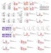
HJURP inhibited PCa cells sensitivity to ferroptosis inducers via the PRDX1/ROS pathway. Increased HJURP levels resulted in increased formation of HJURP-S-S-PRDX1 by direct disulfide binding with PRDX1-SOH or by disulfide bond exchange with PRDX1-S-S-PRDX1, which promoted PRDX1 redox cycling and caused less PRDX1 hyperoxidation. Disulfide binding enhanced the peroxidase activity of PRDX1, leading to a decrease in ROS levels, and ultimately inhibiting the sensitivity of PCa cells to ferroptosis.
figs1
figs1
figs2
figs2
figs3
figs3
figs4
figs4
figs5
figs5
figs6
figs6
figs7
figs7
figs8
figs8

References

    1. Sung H., Ferlay J., Siegel R.L., Laversanne M., Soerjomataram I., Jemal A., et al. Global cancer statistics 2020: GLOBOCAN estimates of incidence and mortality worldwide for 36 cancers in 185 countries. CA A Cancer J. Clin. 2021;71:209–249. - PubMed
    1. Siegel R.L., Miller K.D., Fuchs H.E., Jemal A. Cancer statistics, 2022. CA A Cancer J. Clin. 2022;72:7–33. - PubMed
    1. Takahashi T. Prostate cancer screening and the golden rule of humanity. BMJ. 2023;381:1390. - PubMed
    1. Preisser F., Abrams-Pompe R.S., Stelwagen P.J., Böhmer D., Zattoni F., Magli A., et al. European association of urology biochemical recurrence risk classification as a decision tool for salvage radiotherapy-a multicenter study. Eur. Urol. 2023;85:164–170. - PubMed
    1. Sandhu S., Moore C.M., Chiong E., Beltran H., Bristow R.G., Williams S.G. Prostate cancer. Lancet. 2021;398:1075–1090. - PubMed

Publication types