. 2024 Nov:77:103391.
doi: 10.1016/j.redox.2024.103391. Epub 2024 Oct 9.

Methionine oxidation of actin cytoskeleton attenuates traumatic memory retention via reactivating dendritic spine morphogenesis

Affiliations

Methionine oxidation of actin cytoskeleton attenuates traumatic memory retention via reactivating dendritic spine morphogenesis

Cun-Dong Huang et al. Redox Biol. 2024 Nov.

Abstract

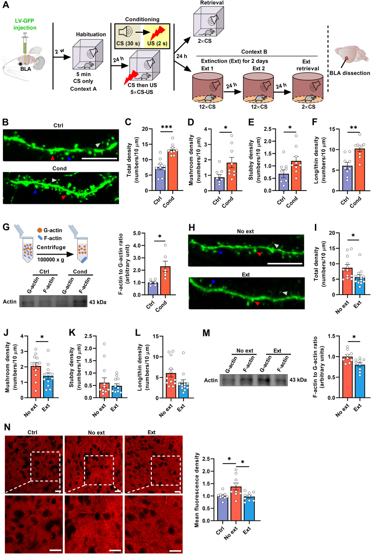
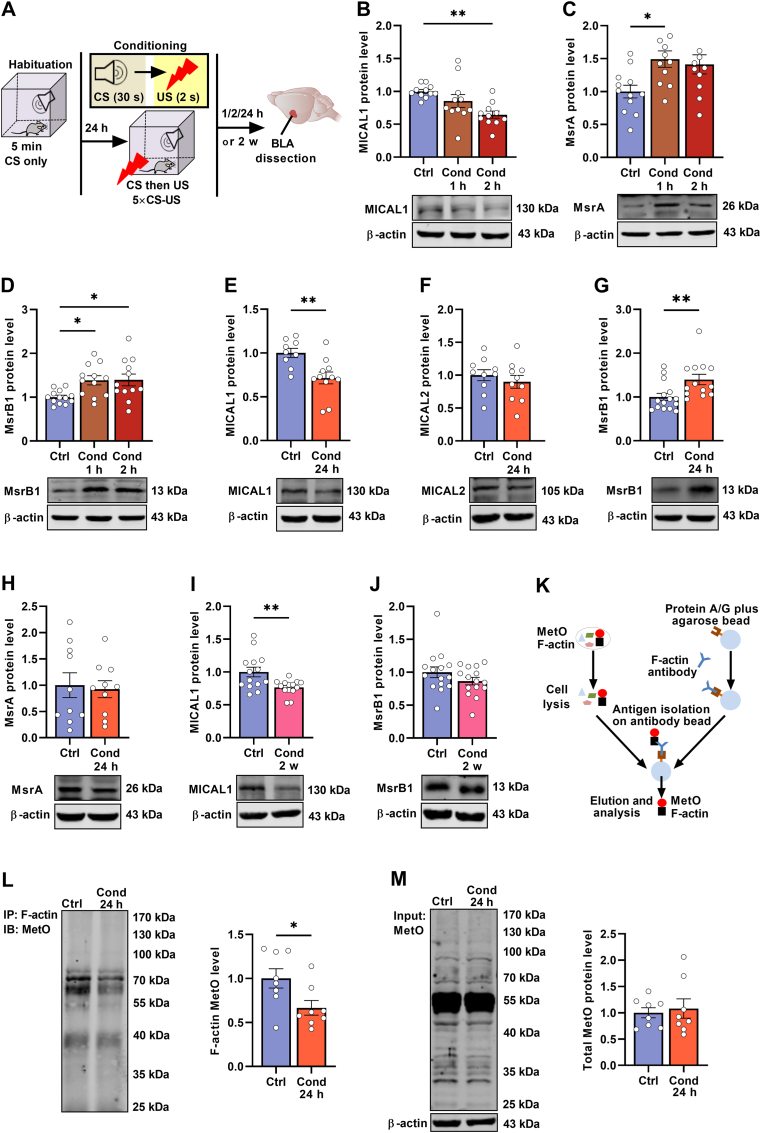
Post-traumatic stress disorder (PTSD) is characterized by hypermnesia of the trauma and a persistent fear response. The molecular mechanisms underlying the retention of traumatic memories remain largely unknown, which hinders the development of more effective treatments. Utilizing auditory fear conditioning, we demonstrate that a redox-dependent dynamic pathway for dendritic spine morphogenesis in the basolateral amygdala (BLA) is crucial for traumatic memory retention. Exposure to a fear-induced event markedly increased the reduction of oxidized filamentous actin (F-actin) and decreased the expression of the molecule interacting with CasL 1 (MICAL1), a methionine-oxidizing enzyme that directly oxidizes and depolymerizes F-actin, leading to cytoskeletal dynamic abnormalities in the BLA, which impairs dendritic spine morphogenesis and contributes to the persistence of fearful memories. Following fear conditioning, overexpression of MICAL1 in the BLA inhibited freezing behavior during fear memory retrieval via reactivating cytokinesis, whereas overexpression of methionine sulfoxide reductase B 1, a key enzyme that reduces oxidized F-actin monomer, increased freezing behavior during retrieval. Notably, intra-BLA injection of semaphorin 3A, an endogenous activator of MICAL1, rapidly disrupted fear memory within a short time window after conditioning. Collectively, our results indicate that redox modulation of actin cytoskeleton in the BLA is functionally linked to fear memory retention and PTSD-like memory.

Keywords: Actin cytoskeleton; Basolateral amygdala; Cued fear conditioning; Dendritic spine; Molecule interacting with CasL 1.


Conflict of interest statement

Declaration of competing interest The authors declare that they have no known competing financial interests or personal relationships that could have appeared to influence the work reported in this paper.

Figures

Image 1
Graphical abstract
Fig. 1
Fig. 1
Fear conditioning induces reversible changes in dendritic spine morphology and cytoskeleton dynamic in BLA. (A) Schematic of the experimental design. (B) Representative images of dendritic spines in the BLA from control and conditioned mice. Scale bars: 10 μm. Blue arrows indicate mushroom-shaped spines, red arrows indicate stubby-shaped spines, and white arrows indicate long/thin-shaped spines. (C-F) Fear conditioning resulted in an increased density of (C) total, (D) mushroom-, (E) stubby- and (F) long/thin-shaped spines in the BLA of conditioned mice (n = 9 neurons from 3 mice per group). (G) The ratio of F-actin to G-actin in the BLA of conditioned mice was significantly elevated (n = 6 mice per group). (H) Representative images of dendritic spines in the BLA of no extinction and extinction mice. Blue arrows: mushroom-shaped spines; Red arrows: stubby-shaped spines; White arrows: long/thin-shaped spines. Scale bars: 10 μm. (I-L) Extinction learning reversed the increases in density of (I) total and (J) mushroom-shaped spines in the BLA, but not (K) stubby- or (L) long/thin-shaped spines (n = 12 neurons from 4 mice per group). (M) Extinction learning decreased the ratio of F-actin to G-actin in the BLA (n = 9 mice per group). (N) Confocal images of F-actin staining using phalloidin in the BLA slices of control, no extinction and extinction mice. Scale bars: 20 μm. Extinction learning resulted in a decreased fluorescence density of F-actin in the BLA (n = 9 slices from 3 mice per group). Ctrl: control; Cond: conditioned; Ext: Extinction; No ext: no extinction. Two-tailed, unpaired t tests (C, D, E, F, J, L, M); unpaired t-test with Welch's correction (G); two-tailed, Mann Whitney tests (I, K); one-way ANOVA with Tukey's multiple comparisons tests (N). ∗p < 0.05, ∗∗p < 0.01 and ∗∗∗p < 0.001. Data are presented as the mean ± SEM. (For interpretation of the references to color in this figure legend, the reader is referred to the Web version of this article.)
Fig. 2
Fig. 2
Fear conditioning decreases the oxidation of actin methionine residues in BLA. (A) Schematic of the experimental design. (B) A significant decrease in protein level of MICAL1 in the BLA of conditioned mice was observed at 2 h post-conditioning (n = 11 mice per group). (C, D) An increased protein level of (C) MsrA and (D) MsrB1 in the BLA of conditioned mice at 1 h post-conditioning (n = 11–12 mice per group). (E-F) At 24 h post-conditioning, a decrease in protein level was noted for (E) MICAL1 in the BLA, while (F) MICAL2 level remained unchanged in conditioned mice (n = 9–11 mice per group). (G) An increase in of MsrB1 protein level was detected in the BLA of conditioned mice at 24 h post-conditioning (n = 13–15 mice per group). (H) The protein level of MsrA in the BLA was showed no significant differences between control and conditioned mice at 24 h post-conditioning (n = 10 mice per group). (I-J) At 2 w post-conditioning, there was a notable decrease in the level of (I) MICAL1, while (J) MsrB1 level remained unchanged in the BLA of conditioned mice (n = 14–15 mice per group). (K) Schematic diagram for illustrating the immunoprecipitation of F-actin interaction with MetO. (L) A decrease in the level of F-actin MetO in the BLA of conditioned mice was observed at 24 h post-conditioning (n = 8 mice per group). (M) No obvious difference in the total level of MetO in the BLA was found at 24 h post-conditioning between control and conditioned mice (n = 8 mice per group). Ctrl: control; Cond: conditioned. One-way ANOVA with Tukey's multiple comparisons tests (B, C, D); two-tailed, unpaired t tests (E, F, H, J, L, M); two-tailed, Mann Whitney tests (G); two-tailed, unpaired t-test with Welch's correction (I); ∗p < 0.05, ∗∗p < 0.01. Data are presented as the mean ± SEM.
Fig. 3
Fig. 3
MICAL1-mediated oxidation of actin in BLA disrupts cued fear memory. (A) Schematic of the experimental design. (B) Schematic representation illustrating the validation of immunohistochemistry for the selective expression of lentivirus constructs in the BLA. Scale bars: 100 μm. (C) The level of F-actin MetO in BLA lysates was significantly increased in the LV-MICAL1 group (n = 8 mice per group). (D) No significant difference in total level of MetO in the BLA was observed between LV-GFP and LV-MICAL1 groups (n = 8 mice per group). (E) The LV-GFP and LV-MICAL1 groups showed equivalent fear acquisition during conditioning (n = 13 mice per group). (F) Overexpression of MICAL1 in the BLA resulted in a decreased freezing behavior on retrieval (n = 19 mice per group). (G, H) The concentrations of (G) H2O2 and (H) MDA in the BLA were similar between the LV-GFP and LV-MICAL1 groups (n = 5–6 mice per group). (I) Schematic representation of the immunohistochemistry validation of the restrictive expression of the lentivirus constructs in the BLA. Scale bars: 100 μm. (J) The LV-scramble and LV-shMsrB1 groups exhibited comparable fear acquisition during conditioning (n = 15–16 mice per group). (K) Knockdown of MsrB1 in the BLA resulted in a decreased freezing level on retrieval (n = 15–16 mice per group). Two-tailed, unpaired t tests (C, D, G, H); two-way ANOVA with Bonferroni's multiple comparisons post hoc tests (E, F, J, K). ∗p < 0.05. Data are presented as the mean ± SEM.
Fig. 4
Fig. 4
Overexpression of MICAL1 in BLA facilitates the extinction of cued fear memory. (A) Behavioral scheme for extinction. (B) Extinction success mice displayed a better performance on extinction retrieval compared to those that experienced extinction failure mice (n = 11–13 mice per group). (C) The expression of MICAL1 in the BLA was found to be upregulated in extinction success mice, (D) whereas MsrB1 did not show similar changes (n = 10–13 mice per group). (E) Schematic of the experimental design. (F) The LV-GFP and LV-MICAL1 groups exhibited an equivalent fear acquisition during conditioning (n = 10–11 mice per group). (G, H) The LV-MICAL1 group showed a better extinction learning on both day 1 and day 2 (n = 10–11 mice per group). (I) The LV-MICAL1 group displayed a significant reduction in freezing behavior on extinction retrieval (n = 10–11 mice per group). (J) Representative fluorescence image for LV-shMICAL1 expression in the BLA. (K) Both of the LV-scramble and LV-shMICAL1 groups showed an equivalent fear acquisition during conditioning (n = 8–9 mice per group). (L, M) The LV-shMICAL1 group showed an increased freezing level during extinction learning on day 2, but not on day 1 (n = 8–9 mice per group). (N) The LV-shMICAL1 group displayed an increased freezing level on extinction retrieval (n = 8–9 mice per group). Ext: Extinction; Ext-success: Extinction success; Ext-failure: Extinction failure; No ext: no extinction. One-way ANOVA with Tukey's multiple comparisons tests (B-D); two-way ANOVA with Bonferroni's multiple comparisons post hoc tests (F, I, K, N); two-way ANOVA with Fisher's LSD tests (G, H, L, M). ∗p < 0.05, ∗∗p < 0.01 and ∗∗∗p < 0.001. Data are presented as the mean ± SEM.
Fig. 5
Fig. 5
Corticosterone-altered MICAL1 expression is involved in the PTSD-like memories. (A) Incubation of HT22 cells with CORT (1 μM) for 24 h resulted in a decreased expression of MICAL1 (n = 6 cells per group). (B) Exposure of CORT (5 mg/kg) over two weeks resulted in a decreased expression of MICAL1 in the BLA of mice (n = 7–8 mice per group). (C, D) No significant differences were observed in the expression levels of (C) MsrA and (D) MsrB1 between vehicle and CORT-exposed mice in the BLA (n = 6–8 mice per group). (E) Schematic of the experimental design. (F) The LV-scramble and LV-shMICAL1 groups showed a similar fear response during conditioning (n = 8 mice per group). (G) Knockdown of MICAL1 in the BLA resulted in an increased freezing behavior on retrieval (n = 16–17 mice per group). (H) Representative fluorescence image from the BLA of the LV-MsrB1 group. (I) The LV-GFP and LV-MsrB1 groups exhibited similar levels of accumulated freezing during conditioning (n = 16 mice per group). (J) Overexpression of MsrB1 in the BLA resulted in an increased freezing behavior on retrieval (n = 14–16 mice per group). (K) Schematic of the experimental design. (L) CORT-exposed mice exhibited an equivalent fear response during conditioning. (M) Overexpression of MICAL1 in the BLA reversed the increased fear response on retrieval in CORT-exposed mice (n = 10 mice per group). CORT: corticosterone. Two-tailed, unpaired t-test with Welch's correction (A, B); two-tailed, unpaired t tests (C, D); two-way ANOVA with Bonferroni's multiple comparisons post hoc tests (F, G, I, J, L, M). ∗p < 0.05, ∗∗p < 0.01. Data are presented as the mean ± SEM.
Fig. 6
Fig. 6
MICAL1-mediated methionine oxidation of actin cytoskeleton reverses fear conditioning-induced increase in dendritic spines in BLA. (A) Schematic of the experimental design. (B) Overexpression of MICAL1 in the BLA decreased the ratio of F-actin to G-actin in conditioned mice (n = 12 mice per group). (C) Overexpression of MICAL1 in the BLA reduced the fluorescence density of F-actin in conditioned mice (n = 9 slices from 3 mice per group). Scale bars: 50 μm. (D) Representative images of dendritic spines from BLA neurons were displayed for the LV-GFP and LV-MICAL1 groups in control and conditioned mice. Scale bars: 10 μm. (E-H) Overexpression of MICAL1 in the BLA reversed the increases in (E) total, (F) mushroom-, and (G) long/thin-shaped spines density in conditioned mice, while having no effect on (H) stubby-shaped spines (n = 9–11 neurons from 3 mice per group). (I) Representative images of dendritic spines in the BLA neurons were obtained from the LV-scramble and LV-shMsrB1 groups of control and conditioned mice. Scale bars: 10 μm. (J-M) Knockdown of MsrB1 in the BLA reversed the increases in (J) total, (K) mushroom-, and (L) long/thin-shaped spines density in conditioned mice, without affecting (M) stubby-shaped spines (n = 10 neurons from 3 mice per group). Ctrl: control; Cond: conditioned. Two-tailed, unpaired t tests (B, C); two-way ANOVA with Bonferroni's multiple comparisons post hoc tests (E-H and J-M). ∗p < 0.05, ∗∗p < 0.01 and ∗∗∗p < 0.001. Data are presented as the mean ± SEM.
Fig. 7
Fig. 7
MICAL1 reactivation in a short window rapidly disrupts cued fear memory. (A) Schematic of the experimental design. (B-E) The effect of MICAL1 reactivation by recombinant mouse SEMA3A on fear memory retrieval was examined. Injection of recombinant mouse SEMA3A into the BLA at (B) 0.5 h, but not at (C) 1 h, (D) 2 h and (E) 24 h post-conditioning, significantly attenuated freezing behavior on retrieval (n = 8 mice per group). Two-way ANOVA with Bonferroni's multiple comparisons post hoc tests (left) or two-tailed, unpaired t tests (right) (B-E). ∗p < 0.05. Data are presented as the mean ± SEM.
Fig. 8
Fig. 8
A working model illustrating how F-actin methionine oxidation dynamics regulates traumatic memories. Traumatic events lead to a down-regulation of MICAL1 expression and an up-regulation of MsrB1 expression in the BLA, resulting in a decrease in F-actin methionine oxidation and an increase in dendritic spine density, which confers long-lasting retention of traumatic memories. Re-activation of MICAL1 counteracts the increase in the actin assembly and dendritic spine complexity in the BLA, thereby inhibiting the retention of traumatic memories.

References

    1. Quirk G.J., Mueller D. Neural mechanisms of extinction learning and retrieval. Neuropsychopharmacology. 2008;33(1):56–72. doi: 10.1038/sj.npp.1301555. - DOI - PMC - PubMed
    1. Furini C., Myskiw J., Izquierdo I. The learning of fear extinction. Neurosci. Biobehav. Rev. 2014;47:670–683. doi: 10.1016/j.neubiorev.2014.10.016. - DOI - PubMed
    1. Maroun M., Ioannides P.J., Bergman K.L., et al. Fear extinction deficits following acute stress associate with increased spine density and dendritic retraction in basolateral amygdala neurons. Eur. J. Neurosci. 2013;38(4):2611–2620. doi: 10.1111/ejn.12259. - DOI - PMC - PubMed
    1. Heinrichs S.C., Leite-Morris K.A., Guy M.D., et al. Dendritic structural plasticity in the basolateral amygdala after fear conditioning and its extinction in mice. Behav. Brain Res. 2013;248:80–84. doi: 10.1016/j.bbr.2013.03.048. - DOI - PMC - PubMed
    1. Tsvetkov E., Carlezon W.A., Benes F.M., et al. Fear conditioning occludes LTP-induced presynaptic enhancement of synaptic transmission in the cortical pathway to the lateral amygdala. Neuron. 2002;34(2):289–300. doi: 10.1016/s0896-6273(02)00645-1. - DOI - PubMed

Publication types

LinkOut - more resources