. 2024:44:103687.
doi: 10.1016/j.nicl.2024.103687. Epub 2024 Oct 11.

Hypothalamic atrophy and structural covariance in amnestic mild cognitive impairment and Alzheimer's dementia

Affiliations

Hypothalamic atrophy and structural covariance in amnestic mild cognitive impairment and Alzheimer's dementia

Hannah Pecher et al. Neuroimage Clin. 2024.

Abstract

Background: Alzheimer's disease (AD) is characterized by progressive cognitive decline and specific brain atrophy patterns, primarily involving the medial temporal lobes. A number of studies have discussed hypothalamic involvement in AD with consecutive metabolic and/or autonomic disturbances yet only few studies have investigated hypothalamic atrophy in AD and its early stages in particular.

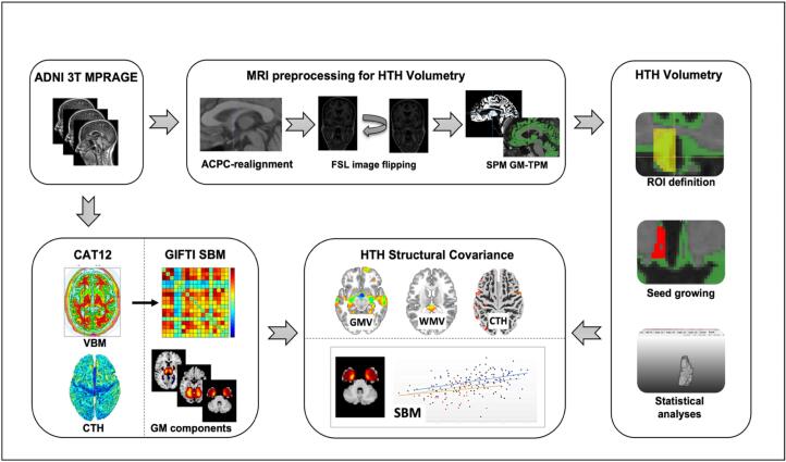
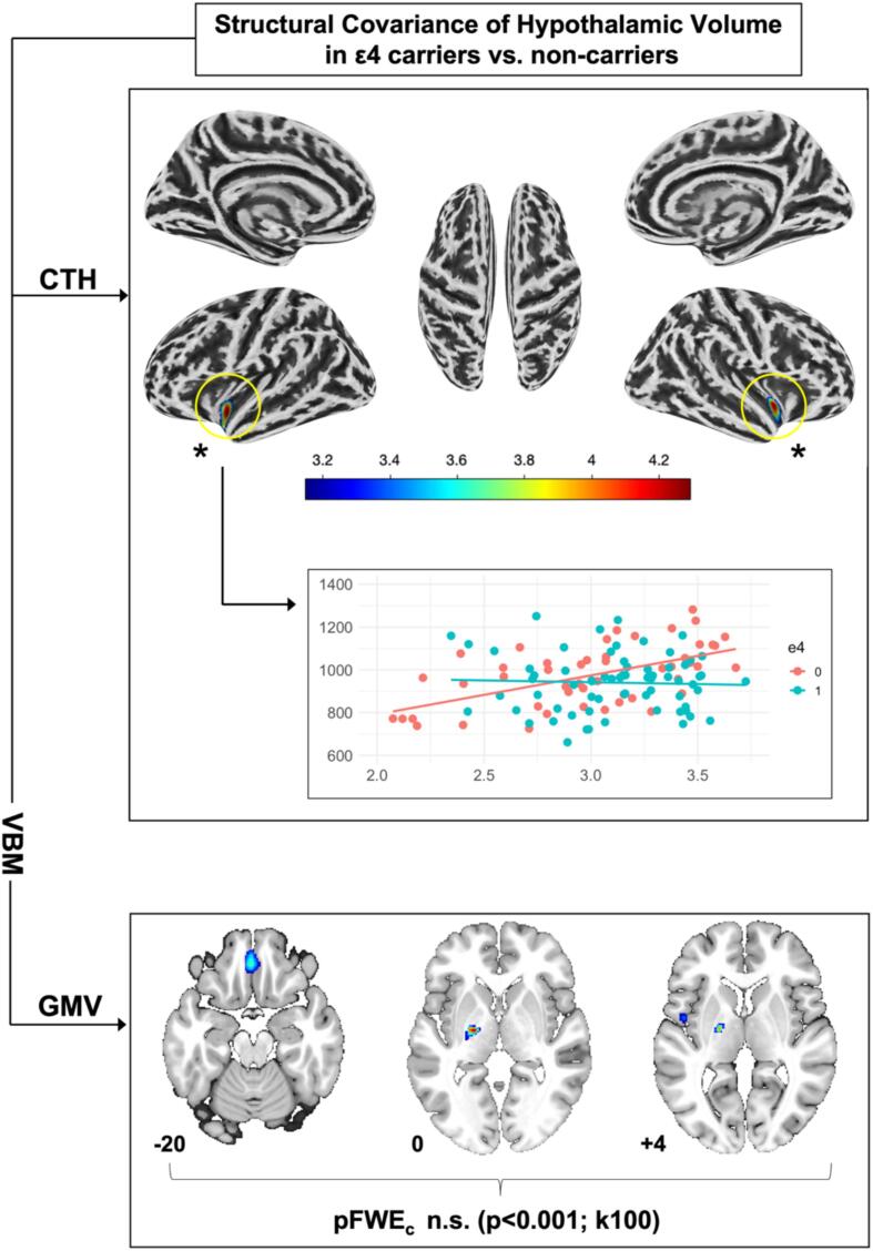
Methods: We applied semi-automated volumetry of the hypothalamus (HTH) in 3 T MRI in a sample N = 175 participants [age 74.9 ± 7.22; gender 85 m/90f; cognitively normal controls (CN; N = 56); amnestic mild cognitive impairment (MCI; N = 78); AD (N = 41)] from the Alzheimer's Disease Neuroimaging Initiative (ADNI). In addition, we used voxel-based morphometry (VBM), cortical thickness (CTH) analyses and source-based morphometry (SBM) derived networks of structural covariance to investigate brain structural covariance patterns of the HTH under consideration of diagnostic groups, β-amyloid (AB) positivity and apolipoprotein E (APOE) ε4 status.

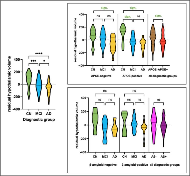
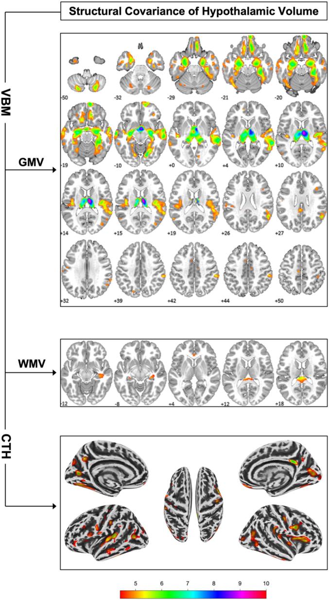
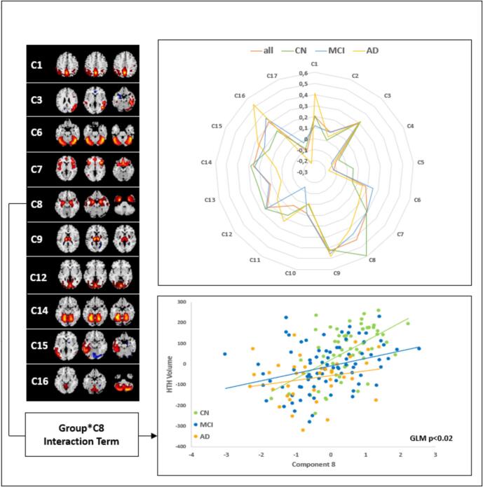
Results: Hypothalamic atrophy was observed in both early and advanced disease stages (i.e. hypothalamic volume CN > MCI > AD). VBM, CTH analysis and SBM revealed positive associations between hypothalamic volume (HV) and AD-vulnerable regions, largely corresponding to the Papez circuit and brain regions implicated in autonomic regulation, however, group differences regarding HTH structural covariance were not observed. Similar observations were made in carriers and non-carriers of the ε4 allele, yet more pronounced in ε4 carriers. Although not reaching significance, comparisons of AB positive vs. negative subjects indicated stronger HTH atrophy in biomarker positive participants. HV was not associated with body mass index or longitudinal weight change.

Conclusions: Our findings support early structural changes of the HTH in AD. HV covaries with regional volumes of AD-vulnerable regions. This could point to secondary atrophy of the HTH following atrophy of the hippocampus and other structures of the Papez circuit in AD.

Keywords: Alzheimer’s disease; Hypothalamic atrophy; Mild cognitive impairment; Structural covariance networks; Volumetry.


Conflict of interest statement

Declaration of competing interest The authors declare that they have no known competing financial interests or personal relationships that could have appeared to influence the work reported in this paper.

Figures

Fig. 1
Fig. 1
Illustrated workflow showing processing of ADNI 3 T MPRAGE data for hypothalamic volumetry as well as structural covariance analysis with SPM12 software and the CAT12 toolbox implemented in SPM12. ADNI Alzheimer’s disease neuroimaging initiative, ACPC anterior commissure posterior commissure, CAT12 computational anatomy toolbox, CTH cortical thickness, GM-TPM gray matter tissue probability map, GMV gray matter volume, HTH hypothalamus, ROI region of interest, SBM source-based morphometry, SPM statistical parametric mapping, VBM voxel-based morphometry, WMV white matter volume.
Fig. 2
Fig. 2
Left: Residualized HV visualised across diagnostic groups showing distribution of HV for cognitively CN subjects, subjects with MCI and AD. Data distribution is visualized using a truncated violin plot with dashed lines indicating median and dotted lines indicating quartiles. Results of pairwise comparison are illustrated with asterisks indicating p-value (ns = P > 0.05; * = P ≤ 0.05; ** = P ≤ 0.01; *** = P ≤ 0.001; **** = P ≤ 0.0001). Right: Residualized HV in APOE- ε4-negative vs. APOE- ε4-positive subjects and β-amyloid-positive and −negative subjects across diagnostic groups with embedded results of post-hoc Duncan’s test showing differences between subgroups. AD alzheimer’s dementia, APOE apolipoprotein E, CN cognitively normal, HV hypothalamic volume, MCI mild cognitive impairment, ns not significant, sign. significant.
Fig. 3
Fig. 3
Structural covariance of hypothalamic volume with gray and white matter volume (i.e. GMV, WMV) via voxel-based morphometry (VBM) and with cortical thickness (CTH). Numbers indicate location on the z axis and colours indicate t-score value. Results are illustrated at p < 0.05 FWE voxel-level corrected and k = 10.
Fig. 4
Fig. 4
Radar chart illustrating the correlations (i.e. Spearman rho) between hypothalamic volume (HV) and individual source-based morphometry (SBM)-based structural covariance networks across diagnostic groups and within the entire sample. Components significantly correlated with HV (i.e. whole sample; pFDR < 0.05) are shown to the left with yellow/red indicating positive covariation and blue colours indicating negative covariation. Scatter plot illustrates the relationship between HV and temporomesial component 8 (C8) and the trend-level significant (p < 0.05 uncorrected) Group*C8 interaction term. HTH hypothalamus. (For interpretation of the references to colour in this figure legend, the reader is referred to the web version of this article.)
Fig. 5
Fig. 5
Structural covariance of hypothalamic volume (HV) with cortical thickness (CTH) and gray matter volume (GMV) depending on apolipoprotein E (APOE) ε4-allele carriership. Numbers indicate location on the z axis and colour indicates t-score value. ε4 = 1 homo- or heterozygous carriership of APOE ε4-allele, ε4 = 0 no APOE ε4-allele carriership. Results of CTH analyses are illustrated at p < 0.05 family-wise error (FWE) voxel-level corrected and k = 10. Voxel-based morphometry (VBM) GMV analyses are illustrated at a liberal, uncorrected threshold of p < 0.001 and k = 100.

References

    1. Aggleton J.P., Nelson A.J.D., O'Mara S.M. Time to retire the serial Papez circuit: Implications for space, memory, and attention. Neuroscience and Biobehavioral Reviews. 2022;140 doi: 10.1016/j.neubiorev.2022.104813. - DOI - PMC - PubMed
    1. Ali, Mohammad; Suh, Jee Su; Ramonas, Milita; Hassel, Stefanie; Arnott, Stephen R.; Strother, Stephen C. et al. (2022): A detailed manual segmentation procedure for the hypothalamus for 3T T1-weighted MRI. In MethodsX 9, p. 101864. DOI: 10.1016/j.mex.2022.101864. - PMC - PubMed
    1. Ashburner J., Friston K.J. Voxel-based morphometry–the methods. NeuroImage. 2000;11(6 Pt 1):805–821. doi: 10.1006/nimg.2000.0582. - DOI - PubMed
    1. Baloyannis S.J., Mavroudis I., Mitilineos D., Baloyannis I.S., Costa V.G. The hypothalamus in Alzheimer's disease: a Golgi and electron microscope study. American Journal of Alzheimer's Disease and Other Dementias. 2015;30(5):478–487. doi: 10.1177/1533317514556876. - DOI - PMC - PubMed
    1. Baloyannis S.J., Mavroudis I., Baloyannis I.S., Costa V.G. Mammillary Bodies in Alzheimer's Disease: A Golgi and Electron Microscope Study. American Journal of Alzheimer's Disease and Other Dementias. 2016;31(3):247–256. doi: 10.1177/1533317515602548. - DOI - PMC - PubMed