. 2024 Nov 6;112(21):3602-3617.e9.
doi: 10.1016/j.neuron.2024.08.021. Epub 2024 Oct 14.

Synaptic neoteny of human cortical neurons requires species-specific balancing of SRGAP2-SYNGAP1 cross-inhibition

Affiliations

Synaptic neoteny of human cortical neurons requires species-specific balancing of SRGAP2-SYNGAP1 cross-inhibition

Baptiste Libé-Philippot et al. Neuron. .

Abstract

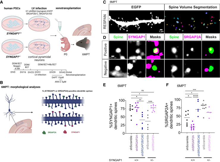
Human-specific (HS) genes have been implicated in brain evolution, but their impact on human neuron development and diseases remains unclear. Here, we study SRGAP2B/C, two HS gene duplications of the ancestral synaptic gene SRGAP2A, in human cortical pyramidal neurons (CPNs) xenotransplanted in the mouse cortex. Downregulation of SRGAP2B/C in human CPNs led to strongly accelerated synaptic development, indicating their requirement for the neoteny that distinguishes human synaptogenesis. SRGAP2B/C genes promoted neoteny by reducing the synaptic levels of SRGAP2A,thereby increasing the postsynaptic accumulation of the SYNGAP1 protein, encoded by a major intellectual disability/autism spectrum disorder (ID/ASD) gene. Combinatorial loss-of-function experiments in vivo revealed that the tempo of synaptogenesis is set by the reciprocal antagonism between SRGAP2A and SYNGAP1, which in human CPNs is tipped toward neoteny by SRGAP2B/C. Thus, HS genes can modify the phenotypic expression of genetic mutations leading to ID/ASD through the regulation of human synaptic neoteny.

Keywords: SRGAP2; SYNGAP1; autism spectrum disorder; cortical neuron; human brain development; intellectual deficiency; neoteny; synapse.


Conflict of interest statement

Declaration of interests The authors declare no competing interests.

Figures

None
Graphical abstract
Figure 1
Figure 1
SRGAP2B/C genes are required for dendritic spine neoteny of human cortical pyramidal neurons (A) Description of the SRGAP2 gene family at the genomic and transcript levels; note the location of the mRNA sequences targeted by shRNAs and that SRGAP2B/C shRNAs do not target the SRGAP2C protein coding sequence (cDNA, in blue). (B) Experimental design of human pluripotent stem cell (PSC)-derived neurons, infected in vitro with lentivirus (LV) expressing EGFP and shRNAs targeting Scramble, SRGAP2B/C, or SRGAP2A sequences ± SRGAP2C-HA cDNA, and then xenotransplanted in the mouse neonatal cerebral cortex followed by morphological analyses (see STAR Methods). (C) Representative proximal dendritic branches of human PSC-derived neurons xenotransplanted in the mouse cerebral cortex, with LV-shRNA infection, followed from 2 to 18 months post-transplantation (MPT). (D) Corresponding quantifications of the dendritic spine density (means ± 95% confident interval; two-way ANOVA tests with Tukey’s multiple comparison test; see Figures S5A and S5D) (numbers in Table S1; n = 12–54 neurons from 2–15 animals from 2–8 litters per stage per condition). (E) Corresponding quantifications of the dendritic spine head width (means ± 95% confident interval; two-way ANOVA tests with Tukey’s multiple comparison test; see Figures S5C and S5E) (numbers in Table S1; n = 151–4,722 dendritic spines from 12–54 neurons from 2–15 animals from 2–8 litters per stage per condition). ∗∗p < 0.01; ∗∗∗∗p < 0.0001. See also Figures S1–S5 and Table S1.
Figure 2
Figure 2
SRGAP2B/C are required for neotenic synaptic maturation of human cortical pyramidal neurons (A) Experimental design of human PSC-derived neurons, LV infected in vitro expressing EGFP and shRNAs targeting Scramble or SRGAP2B/C sequences, and then xenotransplanted in the mouse neonatal cerebral cortex followed by electrophysiological analyses at 6 MPT. (B) Representative traces of synaptic currents of human PSC-derived neurons xenotransplanted in the mouse cerebral cortex, with LV-shRNA infection, at 6 MPT. (C and D) Corresponding quantification of the sEPSCs frequency and amplitude (Mann-Whitney tests) (n = 16–17 neurons per condition from 6–7 animals from 2 litters). (E) Representative examples of AMPA and NMDA currents of human neurons, with LV-shRNA infection, at 6 MPT; the red dashed lines indicate the time points of quantification. (F) Corresponding quantification of the AMPA/NMDA currents amplitude ratio (Mann-Whitney test) (n = 14–19 neurons per condition from 5–7 animals from 2 litters). Data are represented as single neurons with mean + SD. (G and H) Cell capacitance (G) and input resistance (H) of human neurons, with LV-shRNA infection, at 6 MPT (Mann-Whitney test) (n = 16–18 neurons per condition from 6–7 animals from 2 litters). p < 0.05; ∗∗p < 0.01. See also Figure S6.
Figure 3
Figure 3
SRGAP2B/C are human-specific regulators of SRGAP2A/SYNGAP1 antagonism at the synapse in vitro (A) Experimental design: protein extraction at day in vitro 70 (DIV70) from human SYNGAP1+/+ and SYNGAP1+/− PSC-derived neurons with LV-shRNA infection at DIV45. (B) Synaptic fraction of SYNGAP1+/+ and SYNGAP1+/− PSC-derived neurons at DIV70 with LV-shRNA infection at DIV45. The upper and lower parts of the blot (separated by a white lane) were cut prior to primary antibody incubation: the upper part was immunolabeled with antibodies against SYNGAP1, SRGAP2A, and PSD95, and the lower part with antibodies against SRGAP2 and SYP. (SRGAP2B/C) indicates the expected size of SRG2P2B/C, which was not detected in synaptosomes but in the cytosol fraction (see Figure S7C). (C) Corresponding quantifications (ANOVA multiple tests; n = 3–5 experiments per condition). Data are represented as single values with mean + SD. p < 0.05; ∗∗p < 0.01; ∗∗∗p < 0.001; ∗∗∗∗p < 0.0001). See also Figures S7 and S8.
Figure 4
Figure 4
SRGAP2A and SYNGAP1 epistasis regulates synaptic developmental tempo of human cortical pyramidal neurons in vivo (A) Experimental design of SYNGAP1+/+ and SYNGAP1+/− human PSC-derived neurons, LV infected in vitro expressing EGFP and shRNAs targeting Scramble and SRGAP2A sequences, and then xenotransplanted in the mouse neonatal cerebral cortex followed by morphological and electrophysiological analyses at 6 MPT. (B) Representative proximal dendritic branches of SYNGAP1+/+ and SYNGAP1+/− human PSC-derived neurons xenotransplanted in the mouse cerebral cortex, with LV-shRNA infection, at 6 MPT. (C and D) Corresponding quantification of dendritic spine density and spine head width (two-way ANOVA test with Tukey multiple comparison tests) (for SYNGAP1+/+ neurons: n = 30 neurons, 590–980 dendritic spines from 8 animals from 2 litters per condition; for SYNGAP1+/− neurons: n = 30–34 neurons, 1,088–1,433 dendritic spines from 9 animals from 2 litters per condition) (part of SYNGAP1+/+ data are included in Figures 1D and 1E) (see Figure S9A). (E) Corresponding cumulative distribution of spine head width (Kolmogorov-Smirnov tests) (see Figure S9B). (F) Representative examples of synaptic currents of SYNGAP1+/+ and SYNGAP1+/− human PSC-derived neurons xenotransplanted in the mouse cerebral cortex, with LV-shRNA infection, at 6 MPT. (G and H) Corresponding quantifications of sEPSCs frequency and amplitude (Kruskal-Wallis tests with Dunn’s multiple comparison) (for SYNGAP1+/+ neurons: n = 18 neurons from 6 animals from 2 litters; for SYNGAP1+/− neurons: n = 10–23 neurons from 5–6 animals from 3 litters per condition) (SYNGAP1+/+ shScramble data were already shown in Figures 2C and 2D). (I) Cell capacitance of SYNGAP1+/+ and SYNGAP1+/− human PSC-derived neurons xenotransplanted in the mouse cerebral cortex, with LV-shRNA infection, at 6 MPT (Kruskal-Wallis tests with Dunn’s multiple comparison) (for SYNGAP1+/+ neurons: n = 18 neurons from 6 animals from 2 litters; for SYNGAP1+/− neurons: n = 10–23 neurons from 5–6 animals from 3 litters per condition) (SYNGAP1+/+ shScramble data were already shown in Figure 2E). (J) Representative examples of AMPA and NMDA currents of SYNGAP1+/+ and SYNGAP1+/− human PSC-derived neurons xenotransplanted in the mouse cerebral cortex, with LV-shRNA infection, at 6 MPT; the red dashed lines indicate the time points of quantification. (K) Corresponding quantification of the AMPA/NMDA currents amplitude ratio (Kruskal-Wallis tests with Dunn’s multiple comparison) (for SYNGAP1+/+ neurons: n = 15 neurons from 5 animals from 2 litters; for SYNGAP1+/− neurons: n = 10 neurons from 3 animals from 3 litters per condition) (SYNGAP1+/+ shScramble data were already shown in Figure 2G). In (C), (G)–(I), and (K), data are represented as mean + SD. p < 0.05; ∗∗p < 0.01; ∗∗∗∗p < 0.0001. See also Figure S9.
Figure 5
Figure 5
SRGAP2A and SYNGAP1 antagonism at the synapse of human cortical pyramidal neurons in vivo (A and B) Experimental design of SYNGAP1+/+ and SYNGAP1+/− human PSC-derived neurons xenotransplanted in the mouse cerebral cortex, with LV-shRNA infection in vitro expressing EGFP and shRNAs targeting Scramble and SRGAP2A/B/C sequences (A), immunostained at 6 MPT for SYNGAP1 or SRGAP2A and EGFP to quantify the proportion of SYNGAP1-positive and SRGAP2A-positive dendritic spines (B). (C) Representative EGFP dendritic branch from SYNGAP1+/+ shScramble (control) neurons, together with spine head segmentation with RESPAN (see STAR Methods). (D) Representative dendritic spines (single plans) EGFP-positive (green), positive and negative for SRGAP2A or SYNGAP1 (magenta), with corresponding segmentation masks obtained with RESPAN. (E and F) Proportion of SYNGAP1-positive (E) or SRGAP2A-positive (F) dendritic spine per neuron (two-way ANOVA with Tukey’s multiple comparison test; for each condition, n = 4–9 neurons, from 3 animals from 1 litter). Data are represented as single values and median. p < 0.05; ∗∗p < 0.01; ∗∗∗p < 0.001; ∗∗∗∗p < 0.0001.
Figure 6
Figure 6
SRGAP2A and SYNGAP1 epistasis regulates synaptic developmental tempo in mouse cerebral cortex (A) Experimental design of in utero electroporation (IUE) at embryonic day 15.5 (E15.5) that targets the flex-TdTomato construct and Cre recombinase to layer 2/3 CPNs of mouse embryos. Morphometric dendritic spine analysis was then performed on postnatal day (P)21 coronal sections of the following genotypes: control (wild type), Srgap2F/+ (haploinsufficiency), Syngap1F/+ (haploinsufficiency), Srgap2F/+;Syngap1F/+ (dual haploinsufficiency), SRGAP2CKI/+ (gain of function), and Syngap1F/+;SRGAP2CKI/+. (B) Representative apical oblique dendritic branches of mouse L2/3 CPNs at P21 from the respective genotypes. (C) Corresponding quantification of the spine head widths (Kruskal-Wallis tests with Dunn’s multiple comparison) (n = 1,030–2,008 dendritic spines, from 9–15 neurons, from 3–6 mice, from 2–3 litters per condition) (see Figure S10D). (D) Corresponding cumulative distribution of the dendritic spine head widths (Kolmogorov-Smirnov tests) (see Figure S10D). (E and F) Representative western blot (E) and quantification (F) of synaptic abundance of SYNGAP1, SRGAP2(A), and PSD95 in wild-type cerebral cortex at the indicated developmental time points (n = 21 mice from 4 litters, i.e., 3 mice per time point; mean ± SD). ns, not significant. p < 0.05; ∗∗∗∗p < 0.0001. See also Figure S10.

References

    1. DeFelipe J. The Evolution of the Brain, the Human Nature of Cortical Circuits, and Intellectual Creativity. Front. Neuroanat. 2011;5 doi: 10.3389/fnana.2011.00029. - DOI - PMC - PubMed
    1. Sherwood C.C., Gómez-Robles A. Brain Plasticity and Human Evolution. Annu. Rev. Anthropol. 2017;46:399–419. doi: 10.1146/annurev-anthro-102215-100009. - DOI
    1. Vanderhaeghen P., Polleux F. Developmental mechanisms underlying the evolution of human cortical circuits. Nat. Rev. Neurosci. 2023;24:213–232. doi: 10.1038/s41583-023-00675-z. - DOI - PMC - PubMed
    1. Libé-Philippot B., Vanderhaeghen P. Cellular and Molecular Mechanisms Linking Human Cortical Development and Evolution. Annu. Rev. Genet. 2021;55:555–581. doi: 10.1146/annurev-genet-071719-020705. - DOI - PubMed
    1. Wildenberg G., Li H., Sampathkumar V., Sorokina A., Kasthuri N. Isochronic development of cortical synapses in primates and mice. Nat. Commun. 2023;14 doi: 10.1038/s41467-023-43088-3. - DOI - PMC - PubMed

Substances

LinkOut - more resources