. 2025 Jan;17(1):82-93.
doi: 10.1111/os.14260. Epub 2024 Oct 15.

A More Efficient and Safer Improved Percutaneous Pedicle Screw Insertion Technique-Trajectory Dynamic Adjustment Technique, Technical Note, and Clinical Efficacy

Affiliations

A More Efficient and Safer Improved Percutaneous Pedicle Screw Insertion Technique-Trajectory Dynamic Adjustment Technique, Technical Note, and Clinical Efficacy

Hao Li et al. Orthop Surg. 2025 Jan.

Abstract

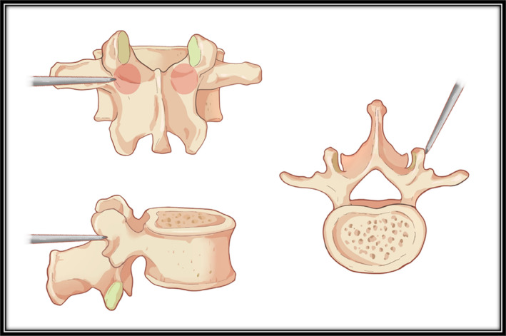
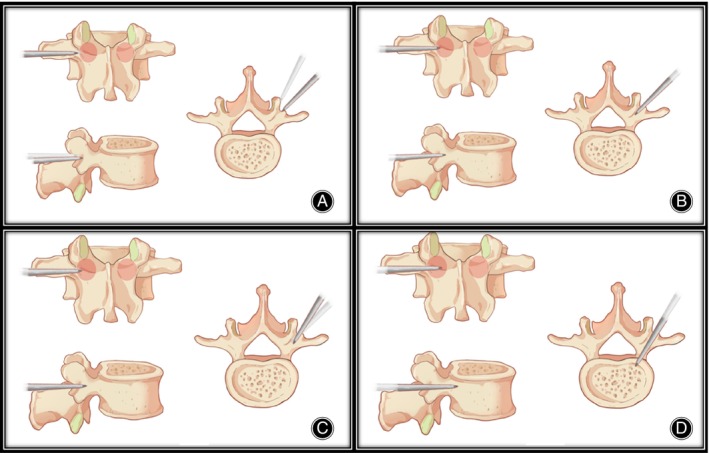
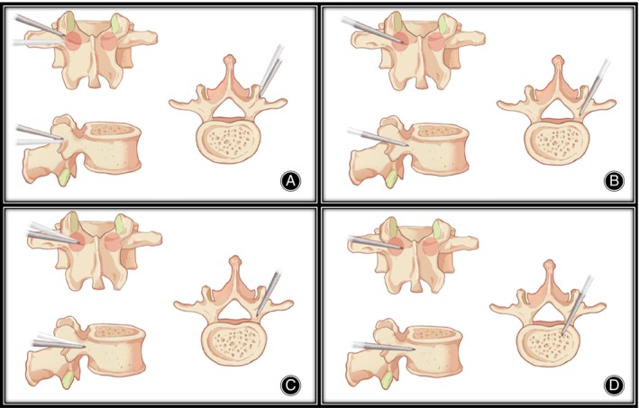
Objective: Percutaneous pedicle screw fixation (PPSF) technique requires a very precise entry point of the Jamshidi needle, which leads to repeated adjustments, damaging the pedicle and increasing radiation exposure. This study was designed to propose an improved percutaneous pedicle screw fixation technique-trajectory dynamic adjustment (TDA) technique, and evaluate its feasibility and assess the clinical outcomes.

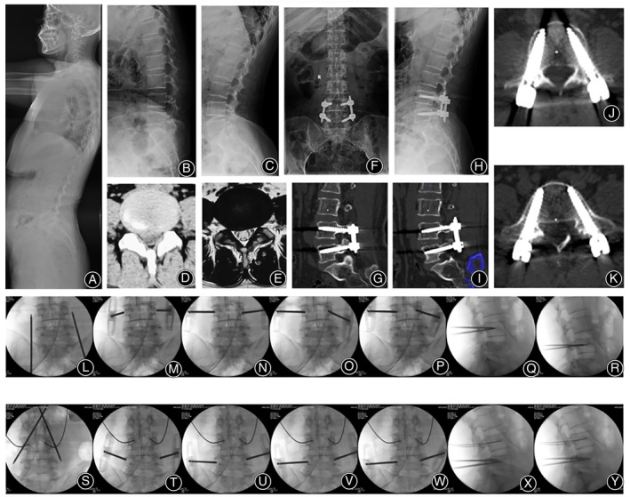
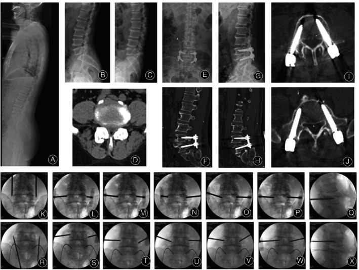
Method: A total of 445 patients with lumbar spondylolisthesis or lumbar spinal stenosis associated with instability from June 2017 to May 2022 were included in the retrospective study. They were randomly separated into two groups. Two hundred thirty-one patients underwent TDA technique (TDA group). Two hundred fourteen patients underwent traditional PPSF technique (PPSF group). All patients underwent postoperative CT to assess the accuracy of screw placement, superior facet joint violation (FJV). The evaluated clinical outcomes were needle insertion time, radiation exposure, blood loss, hospital stay, the Japanese Orthopedic Association (JOA) score, the Visual Analogue Scale (VAS) scores for lower back pain (LBP), and leg pain, lumbar interbody fusion rate, and postoperative complications. The independent-sample t test and paired t-test were used for continuous data. The contingency table and Mann-Whitney U test were used for categorical data.

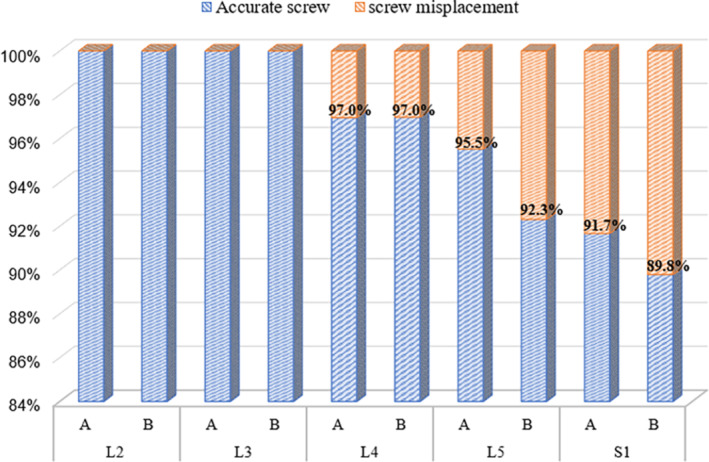
Results: The time of the insertion in TDA group was significantly lower than that in PPSF group (p < 0.05). Similarly, the fluoroscopy frequency in TDA group was significantly lower than that in PPSF group (p < 0.05). There was no difference in intraoperative blood loss and hospital stay between the two groups (p > 0.05). Overall, there was no significant difference in the proportion of clinically acceptable screws between the two groups (p > 0.05). In addition, the lateral screw misplacement in TDA group was higher. Moreover, FJV rate was significantly lower than that in PPSF group (p < 0.05). In both TDA group and PPSF group, postoperative back and leg pain and the JOA score were significantly improved (p < 0.05). However, there were no significant differences in the pre- and postoperative VAS score for back and leg pain and the JOA score, JOA recovery rate, intervertebral fusion rate, and complications rate between the two groups (p > 0.05).

Conclusion: Compared to traditional PPSF technique, TDA technique is a safer and more effective procedure which has shorter surgical time, lower radiation exposure, and lower facet joint violation rate.

Keywords: facet joint violation; lumbar; minimally invasive; percutaneous pedicle screw fixation; radiation exposure.


Conflict of interest statement

The authors declare no conflicts of interest.

Figures

FIGURE 1
FIGURE 1
Entry point of traditional percutaneous pedicle screw fixation.
FIGURE 2
FIGURE 2
The process of coronal/axial trajectory adjustment. (A) The entry point is more outward than a traditional PPSF, and lateral tilt angle of the needle can be appropriately increased. (B) The needle tip enters the pedicle. (C) Reduce the lateral tilt angle of the needle. (D) The needle tip enters the vertebral body.
FIGURE 3
FIGURE 3
The process of sagittal trajectory adjustment. (A) The entry point is outside and above compared with traditional PPSF, the head tilt angle of the needle should be appropriately increased. (B) The needle tip gradually approached to the midline of the pedicle. (C) Reduce the head tilt angle of the needle. (D) The needle tip enters the vertebral body.
FIGURE 4
FIGURE 4
A 65‐year‐old female patient was diagnosed with degenerative spondylolisthesis, and MIS‐TLIF with TDA technique was performed. (A–C) Preoperative x‐rays showed instability of L4‐5. CT (D) and MRI (E) showed disc herniation. (F–K) Postoperative x‐rays and CT showed accurate screw placement. Figure (L) shows the localization of the L4 puncture points. The initial anchoring point on both sides was located 3–5 mm outside the lateral wall of the pedicle, with a slightly smaller angle of puncture needles on both sides (M). After increasing the abduction angle on the left side, the needle tip advanced to the medial wall of the pedicle, which was the ideal path for the screw; meanwhile, the puncture point on the right side slid down to the lower margin of the pedicle, representing a non‐ideal path (N). The left puncture needle maintained its direction and advanced to the inner wall of the pedicle, while the right puncture needle tilted caudally, directing the needle tip toward the cephalad and gradually advancing to the center of the upper and lower margins of the pedicle (O). The right puncture needle continued to advance until the needle tip reached the inner wall of the pedicle (P). Lateral fluoroscopy showed that the needle tips had entered the vertebral body (Q). The needle cores were removed, and guide wires were placed (R). Figure S illustrates the localization of the L5 puncture points. The initial anchoring point on the left side was ideal, while the right side was deviated toward the head side, with a smaller abduction angle (T). When advancing the puncture needle on the left side, it slid caudally to a non‐ideal position, while the needle of right side continued to advance with an increased abduction angle and head tilt angle (U). The left puncture needle tilted caudally, directing the needle tip to the center of the upper and lower margins of the pedicle, and continued to advance; the right side maintained the original angle while continuing to advance (V). Needles of both sides continued to advance until the needle tips reached the inner wall of the pedicle (W). At this point, lateral fluoroscopy showed that the needle tips had entered the vertebral body (X). The needle cores were removed, and guide wires were placed (Y).
FIGURE 5
FIGURE 5
A 60‐year‐old male patient was diagnosed with lumbar spinal stenosis associated with instability, and MIS‐TLIF with TDA technique was performed. (A–C) Preoperative x‐rays showed instability of L4‐5. CT (D) showed lumbar stenosis. (E–J) Postoperative x‐rays and CT showed accurate screw placement. Figure (K) illustrated the positioning of the L5 puncture point. The initial anchoring point on the left side was in a less‐than‐ideal position, leaning toward the head; the initial anchoring point on the right side was slightly external (L). The position of the left anchoring point was not adjusted, with a moderate increase in the head tilt direction, directing the needle tip toward the center of the upper and lower edges of the pedicle; the right anchoring point was adjusted inward, remaining in a less than ideal position, also leaning toward the head side, with a smaller abduction angle (M). The lateral abduction angle was increased, and the head tilt angle was also increased, allowing the needle tip to reach the inner wall of the pedicle; this operation caused the skin to become overly tight, resulting in a smaller abduction angle of the left puncture needle (N). At this point, the right puncture needle had achieved a secure anchoring, prompting an increase in the left puncture needle's abduction angle to continue advancing the needle (O). Slight adjustments were made to both sides of the puncture needles, and the advancement continued until the needle tip reached the inner wall of the pedicle (P). At this moment, the lateral view showed that the needle tips had entered the vertebral body (Q). Figure R illustrated the positioning of the L4 puncture point. The initial anchoring point on the left side leaned toward the head, while the right side leaned toward the tail (S). During the adjustment of the anchoring points, the left side leaned toward the tail, and the right side leaned toward the head (T and U). During the advancement of the needle, the left side increased its tail tilt, while the right side increased its head tilt, allowing the needle tip to reach the inner wall of the pedicle (U and W). At this point, the lateral view showed that the needle tips had entered the vertebral body (X).
FIGURE 6
FIGURE 6
Clinically acceptable screws in different segments between two groups.

Similar articles

Cited by

References

    1. Magerl F., “External Skeletal Fixation of the Lower Thoracic and the Lumbar Spine,” in Current Concepts of External Fixation of Fractures, eds. Uhthoff H. K. and Stahl E. (Berlin, Heidelberg: Springer Berlin Heidelberg, 1982), 353–366.
    1. Mathews H. H. and Long B. H., “Minimally Invasive Techniques for the Treatment of Intervertebral Disk Herniation,” Journal of the American Academy of Orthopaedic Surgeons 10 (2002): 80–85, 10.5435/00124635-200203000-00003. - DOI - PubMed
    1. Foley K. T., Gupta S. K., Justis J. R., and Sherman M. C., “Percutaneous Pedicle Screw Fixation of the Lumbar Spine,” Neurosurgical Focus 10 (2001): E10–E19, 10.3171/foc.2001.10.4.11. - DOI - PubMed
    1. Ishii K., Funao H., Isogai N., et al., “The History and Development of the Percutaneous Pedicle Screw (PPS) System,” Medicina 58 (2022): 1064, 10.3390/medicina58081064. - DOI - PMC - PubMed
    1. Anand N., Baron E. M., Thaiyananthan G., Khalsa K., and Goldstein T. B., “Minimally Invasive Multilevel Percutaneous Correction and Fusion for Adult Lumbar Degenerative Scoliosis: A Technique and Feasibility Study,” Journal of Spinal Disorders & Techniques 21 (2008): 459–467, 10.1097/BSD.0b013e318167b06b. - DOI - PubMed