. 2024 Oct 15;15(1):8783.
doi: 10.1038/s41467-024-52726-3.

Identification of Shemin pathway genes for tetrapyrrole biosynthesis in bacteriophage sequences from aquatic environments

Affiliations

Identification of Shemin pathway genes for tetrapyrrole biosynthesis in bacteriophage sequences from aquatic environments

Helen Wegner et al. Nat Commun. .

Abstract

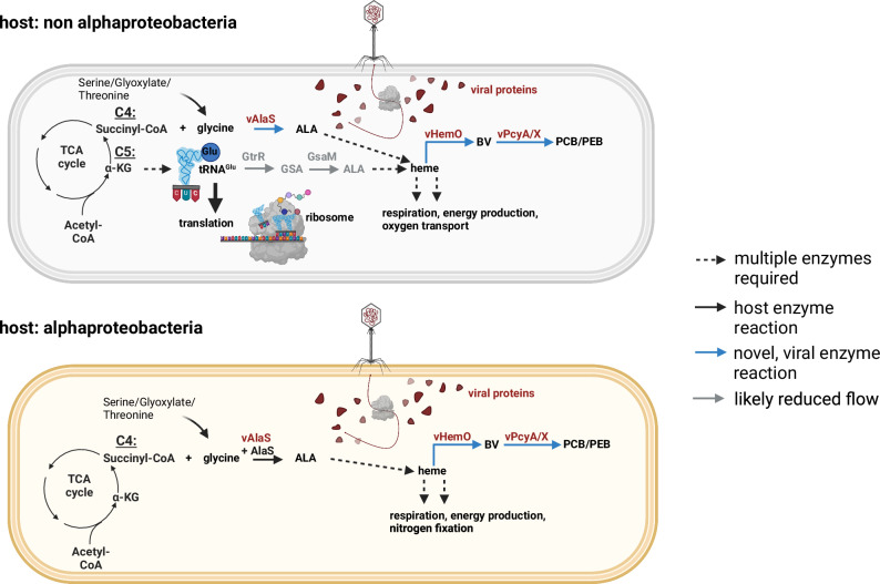
Tetrapyrroles such as heme, chlorophyll, and vitamin B12 are essential for various metabolic pathways. They derive from 5-aminolevulinic acid (5-ALA), which can be synthesized by a single enzyme (5-ALA synthase or AlaS, Shemin pathway) or by a two-enzyme pathway. The genomes of some bacteriophages from aquatic environments carry various tetrapyrrole biosynthesis genes. Here, we analyze available metagenomic datasets and identify alaS homologs (viral alaS, or valaS) in sequences corresponding to marine and freshwater phages. The genes are found individually or as part of complete or truncated three-gene loci encoding heme-catabolizing enzymes. Amino-acid sequence alignments and three-dimensional structure prediction support that the valaS sequences likely encode functional enzymes. Indeed, we demonstrate that is the case for a freshwater phage valaS sequence, as it can complement an Escherichia coli 5-ALA auxotroph, and an E. coli strain overexpressing the gene converts the typical AlaS substrates glycine and succinyl-CoA into 5-ALA. Thus, our work identifies valaS as an auxiliary metabolic gene in phage sequences from aquatic environments, further supporting the importance of tetrapyrrole metabolism in bacteriophage biology.


Conflict of interest statement

The authors declare no competing interests.

Figures

Fig. 1
Fig. 1. Tetrapyrrole biosynthesis pathway.
The common precursor 5-aminolevulinic acid (5-ALA) is synthesized by two different pathways. While plants, most bacteria and archaea use the C5 pathway employing glutamyl t-RNA reductase (GtrR) and glutamate-1-semialdehyde-2,1-aminomutase (GsaM), the aminolevulinic acid synthase (AlaS) within the Shemin pathway of alphaproteobacteria, mammals and birds condenses glycine and succinyl-CoA to yield 5-ALA. Eight molecules of 5-ALA are needed to synthesize intermediate uroporphyrinogen III, an intermediate to most modified tetrapyrroles. Heme can then be converted through heme oxygenase (HemO) to linear biliverdin IXα, which can be converted through ferredoxin-dependent bilin reductases to phycocyanobilin (PCB) or phycoerythrobilin (PEB). PcyA=phycocyanobilin:ferredoxin oxidoreductase; PebS/PcyX=phycoerythrobilin synthase; DHBV=dihydrobiliverdin. The side chains are abbreviated as follows: P=propionate; A=acetate; M=methyl; V=vinyl. Created with parts from BioRender.com, released under a Creative Commons Attribution-NonCommercial-NoDerivs 4.0 International license.
Fig. 2
Fig. 2. Genomic contigs containing valaS.
Schematic representation of representative genomic contigs retrieved in this project. Genes are colored by categories according to the genome legend. Gene annotations are written within the gene when available; “gene products (gp)” are marked solely by their number. Contigs longer than presented are marked with a dotted line. The scale bar denotes bp. Sequences for the contigs can be found in Supplementary Data 4.
Fig. 3
Fig. 3. Phylogenetic analyzes of AlaS protein sequences from bacterial and viral origin.
Unrooted maximum likelihood phylogenetic tree for AlaS. Black names denote sequences from bacteria and metagenomic contigs of uncertain origin. Bradyrhizobium sp. ORS278, containing the three gene-cassette, is marked in gold. The vAlaS sequence from CB_2 phage chosen for experimental characterization is colored pink. Metagenomically retrieved contigs from this project are color coded according to the bars in Fig. 2. Circles represent bootstrap values > 0.9. The scale bar indicates the average number of amino acid substitutions per site.
Fig. 4
Fig. 4. Structural model of monomer vAlaS (CB_2) (red) and overlay with bacterial AlaS (blue) from R. capsulatus using AlphaFold.
The dimer structure of vAlaS is depicted transparently in conjunction with its second monomer. Yellow spheres represent the pyridoxal phosphate (PLP) cofactor, one on each monomer. Glycine binding site (green circle) and succinyl-CoA binding site (purple circle). The lower picture shows an enhanced view of the PLP binding site where a Schiff’s base is formed with amino acid residue K248 (R. capsulatus numbering). A PDB file of the vAlaS modeled structure is available as Supplementary Data 7.
Fig. 5
Fig. 5. Functional complementation of ALA-auxotrophic E. coli ST18 with a functional and non-functional vAlaS.
a. Growth of ST18 containing plasmid encoded vAlaS (squares), plasmid encoded vAlaSK250A variant (diamond) and the vector control (circles) in the presence (filled) or absence (empty symbol) of inductor IPTG. The data are mean values for three biological replicates for each strain and condition. Standard deviation is shown in error bars at each time point. b. vAlaS shows distinct enzyme activity in cell lysate when supplemented with C4 specific substrates. Cell lysate of complemented E. coli strains was supplemented with glycine and succinyl‑CoA to detect product formation over time. Time points demonstrate the respective amount of ALA that was measured after subtracting the already present ALA in the cell (time point 0 min). The data shown represents the mean values of all measurements and the respective standard deviation. Measurements were conducted with three independent biological replicates and two technical replicates (n = 6). For statistical determination, values were tested for a normal distribution using the Shapiro‒Wilk test, and normally distributed data were analyzed using one-way ANOVA with Bonferroni correction and paired t tests to determine the significance levels of each time point against time point 0. Significance levels were assigned accordingly: p ≤ 0.05 equals *; p ≤ 0.01 equals ** and p ≤ 0.001 equals ***. Source data are provided as a Source Data file.
Fig. 6
Fig. 6. Enzyme activity for the vHemO- and vPcyA-catalyzed reactions.
a HPLC analyzes of bilin reductase and heme oxygenase activity monitored at 650 nm. One reaction product of vPcyA is marked with an asterisk and could be the intermediate 181,182-DHBV. BphO, heme oxygenase; PcyA, cyanobacterial PCB:ferredoxin oxidoreductase. b Phytochrome difference spectrum was obtained following coexpression of genes encoding Cph1, vHemO and vPcyA and purification of Cph1 via metal affinity chromatography. In vivo chromophore assembly was detected after illumination with red light (636 nm—Pfr spectrum) and far-red light (730 nm for Cph1—Pr spectrum). Difference spectra were calculated by subtracting Pfr from the Pr spectrum. Source data are provided as a Source Data file.
Fig. 7
Fig. 7. Proposed model of phages carrying all three tetrapyrrole biosynthesis genes and their impact on alphaproteobacterial and non-alphaproteobacterial cells.
We hypothesize that in a non alphaprotobacterial host vALAS is responsible for tetrapyrrole biosynthesis during infection, enabling the virocell to employ glutamyl-tRNA for protein biosynthesis instead of tetrapyrrole biosynthesis. In alphaproteobacterial hosts, we propose that vAlaS supports its own AlaS in help of generating sufficient amounts of tetrapyrroles to support respiration and energy production. See text for more details. Created with BioRender.com, released under a Creative Commons Attribution-NonCommercial-NoDerivs 4.0 International license.

References

    1. Battersby, A. R. Tetrapyrroles: the pigments of life. Nat. Prod. Rep.17, 507–526 (2000). - PubMed
    1. Bryant, D. A., Hunter, C. N. & Warren, M. J. Biosynthesis of the modified tetrapyrroles-the pigments of life. J. Biol. Chem.295, 6888–6925 (2020). - PMC - PubMed
    1. Shemin, D. & Rittenberg, D. The utilization of glycine for the synthesis of a porphyrin. J. Biol. Chem.159, 567–568 (1945).
    1. Beale, S. & Castelfranco, P. 14C incorporation from exogenous compounds into delta-aminolevulinic acid by greening cucumber cotyledons. Biochem. Biophys. Res. Commun.52, 143–149 (1973). - PubMed
    1. Dammeyer, T., Bagby, S. C., Sullivan, M. B., Chisholm, S. W. & Frankenberg-Dinkel, N. Efficient phage-mediated pigment biosynthesis in oceanic cyanobacteria. Curr. Biol.18, 442–448 (2008). - PubMed

Publication types

MeSH terms

Associated data

LinkOut - more resources