. 2024 Oct 15;15(1):8887.
doi: 10.1038/s41467-024-53277-3.

The subcortical maternal complex modulates the cell cycle during early mammalian embryogenesis via 14-3-3

Affiliations

The subcortical maternal complex modulates the cell cycle during early mammalian embryogenesis via 14-3-3

Zhuo Han et al. Nat Commun. .

Abstract

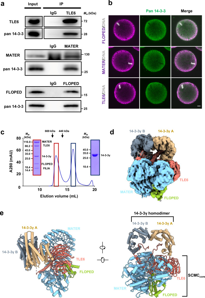
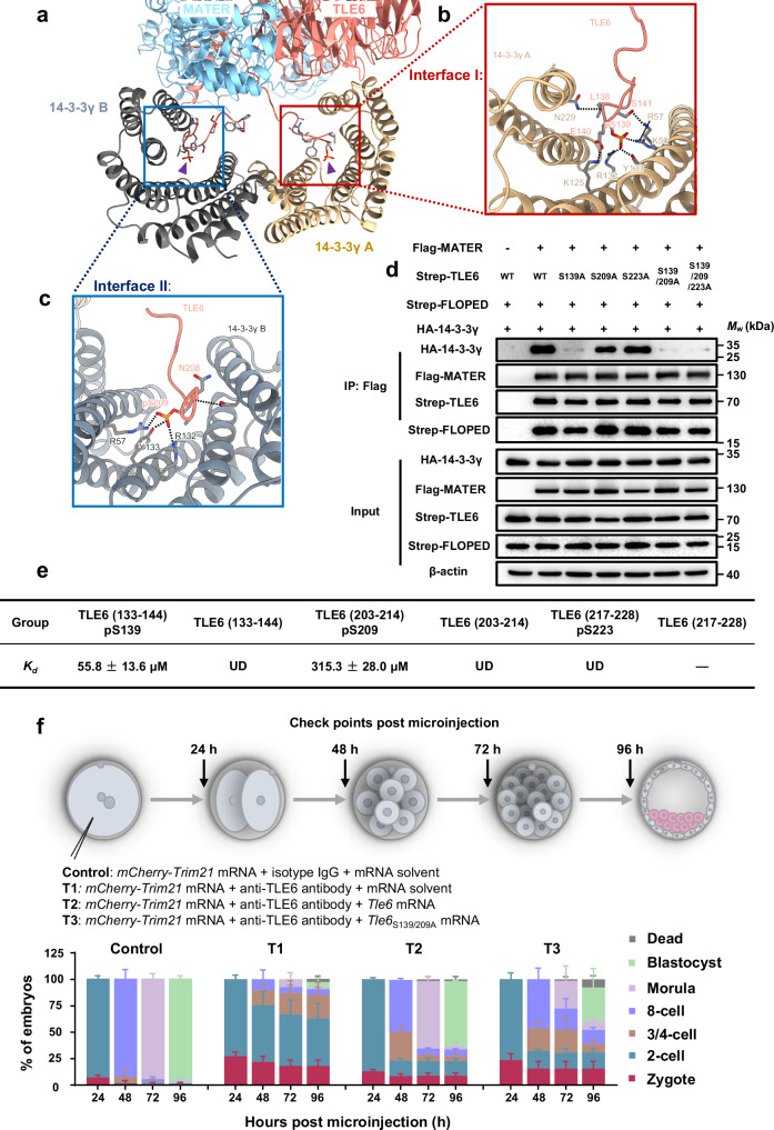
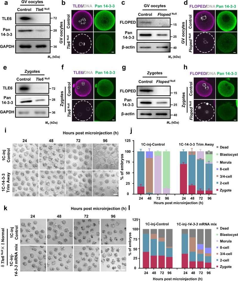
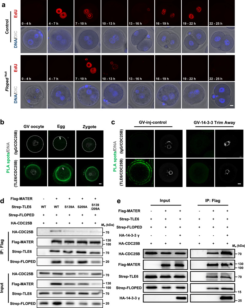
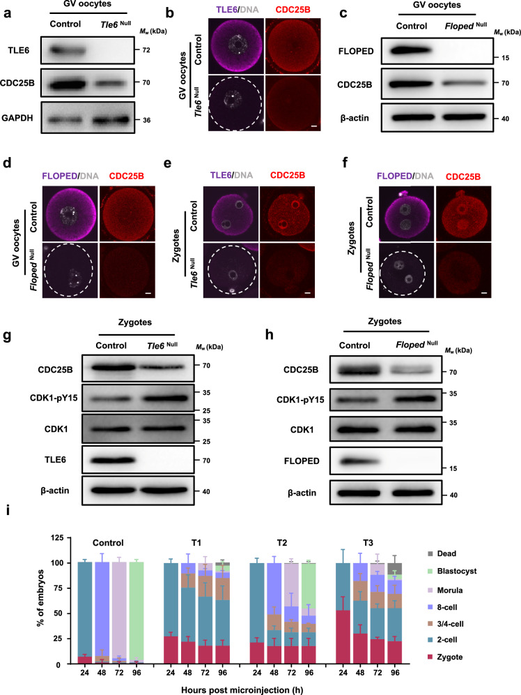
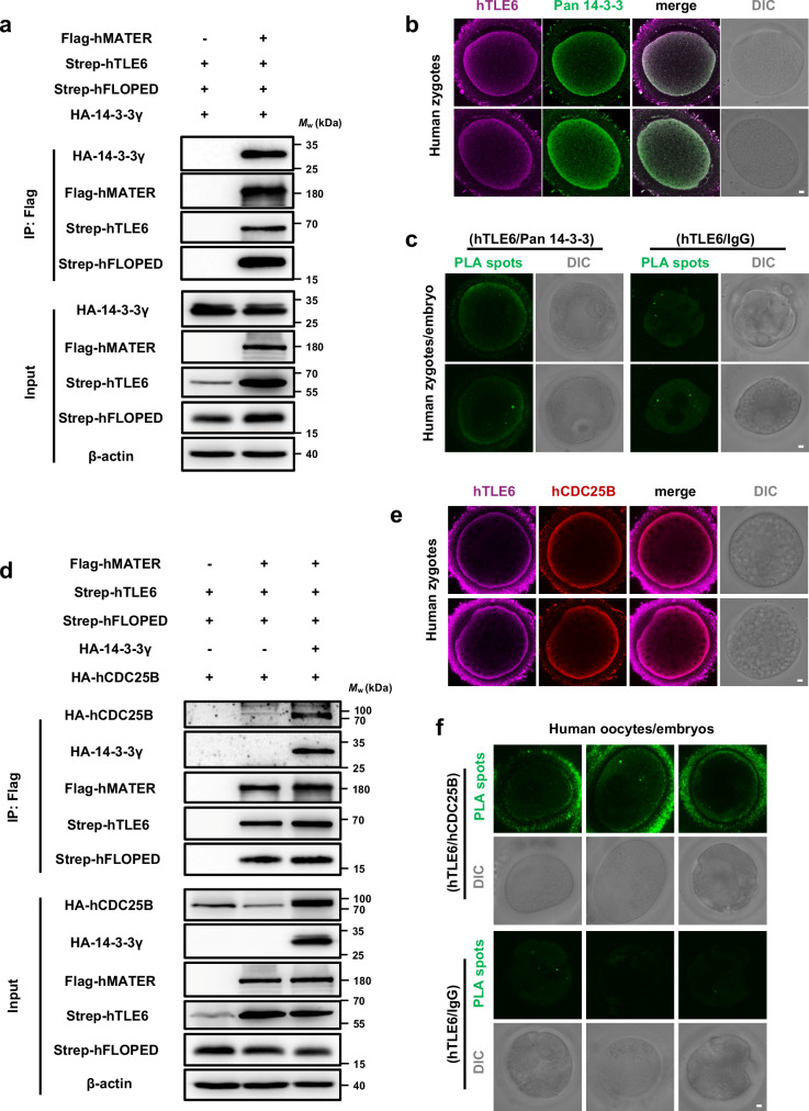
The subcortical maternal complex (SCMC) is essential for safeguarding female fertility in mammals. Assembled in oocytes, the SCMC maintains the cleavage of early embryos, but the underlying mechanism remains unclear. Here, we report that 14-3-3, a multifunctional protein, is a component of the SCMC. By resolving the structure of the 14-3-3-containing SCMC, we discover that phosphorylation of TLE6 contributes to the recruitment of 14-3-3. Mechanistically, during maternal-to-embryo transition, the SCMC stabilizes 14-3-3 protein and contributes to the proper control of CDC25B, thus ensuring the activation of the maturation-promoting factor and mitotic entry in mouse zygotes. Notably, the SCMC establishes a conserved molecular link with 14-3-3 and CDC25B in human oocytes/embryos. This study discloses the molecular mechanism through which the SCMC regulates the cell cycle in early embryos and elucidates the function of the SCMC in mammalian early embryogenesis.


Conflict of interest statement

The authors declare no competing interests.

Figures

Fig. 1
Fig. 1. 14-3-3 exhibits as an SCMC component.
a Normal oocyte lysates, before (Input) or after immunoprecipitation with antibody to TLE6 (top), MATER (middle) or FLOPED (bottom) were immunoblotted and probed with antibodies to pan 14-3-3 and TLE6, MATER, or FLOPED, respectively. IgG, normal immunoglobulin (negative control). b Normal oocytes isolated from normal females were fixed, permeabilized, and incubated with antibodies to pan 14-3-3 and FLOPED (top), MATER (middle), or TLE6 (bottom), and with DAPI to visualize DNA. The oocytes were imaged by confocal microscopy. Scale bar, 10 μm. Colocalization of pan 14-3-3 and FLOPED, MATER, or TLE6 was presented in the merge. c In vitro reconstitution of mouse SCMC-14-3-3γ. The mouse SCMC quaternary complex and 14-3-3γ protein were incubated in lysis buffer. Size-exclusion chromatography (Superose™ 6 Increase 10/300 GL) was performed to separate the SCMC-14-3-3γ (marked in dark red box) and excess 14-3-3γ (marked in dark blue box). The SCMC-14-3-3γ is composed of His-tagged MATER (1–1059 aa), Strep-tagged TLE6 (48–581 aa), Strep-tagged FLOPED (1–164 aa), Strep-tagged FILIA (1–124 aa), and 14-3-3γ (1–247 aa). The column was calibrated with thyroglobulin (669 kDa) and ferritin (440 kDa). d Cryo-EM map of the SCMC-14-3-3γ. 14-3-3γ A and 14-3-3γ B refer to two protomers in 14-3-3γ homodimer. MATER, TLE6, and FLOPED are colored in light blue, salmon, and yellow green, respectively. The homodimer of 14-3-3γ is distinguished by 14-3-3γ A (burlywood), which is proximal to the WD40 repeat domain of TLE6, and 14-3-3γ B (gray), which is close to the leucine-rich repeat domain of MATER. The map was shown at level 0.00679. e Two views of the cartoon presentation of the SCMC-14-3-3γ structure. The color scheme is consistent with panel d. The SCMCcore indicates the core complex consisting of MATER, TLE6, and FLOPED.
Fig. 2
Fig. 2. Site-specific phosphorylation of TLE6 is important for binding 14-3-3.
ac Interface I and II mediating the binding of two 14-3-3γ protomers to TLE6 in the SCMC. Interface I (b, marked in red box) and Interface II (c, marked in blue box) show the interactions of two 14-3-3γ protomers with TLE6 pS139 and pS209, respectively. Purple triangles indicate TLE6 pS139 and pS209, respectively. pS, phosphorylated serine. Single components are colored and labeled as denoted. d HEK-293F cells were co-transfected with expression vectors harboring Flag-tagged MATER (or a blank vector for negative control), Strep-tagged FLOPED, HA-tagged 14-3-3γ and either Strep-tagged TLE6, TLE6S139A mutant, TLE6S209A mutant, TLE6S223A mutant, TLE6S139/209A double-site mutant, or TLE6S139/209/223A triple-site mutant as denoted. 60 h after transfection, cell lysates before (Input) or after immunoprecipitation with anti-Flag affinity agarose gel were immunoblotted for HA-tagged 14-3-3γ, Flag-tagged MATER, Strep-tagged FLOPED and Strep-tagged TLE6 (or mutants). e Table for binding affinity (Kd) between His-tagged 14-3-3γ and TLE6 wildtype peptides (TLE6 (133–144) or TLE6 (203–214)) or phosphorylated peptides (TLE6 (133–144) pS139, TLE6 (203–214) pS209, or TLE6 (217–228) pS223). The data represent the mean ± S.D. (standard deviation) of 3 independent experiments. pS, phosphorylated serine. UD, undetectable. The minus symbol “-” indicates that the peptide TLE6 (217–228) was undetermined due to insolubility. f Schematic overview of mouse zygotes cultured for 24, 48, 72, and 96 h after co-microinjection with mCherry-Trim21 mRNA + isotype IgG + mRNA solvent (control), mCherry-Trim21 mRNA + anti-TLE6 antibody + mRNA solvent (T1), mCherry-Trim21 mRNA + anti-TLE6 antibody + Tle6 mRNA (T2), or mCherry-Trim21 mRNA + anti-TLE6 antibody + Tle6S139/209A mRNA (T3). The validation for the loss of TLE6 was presented in Supplementary Fig. 7b. The proportions of embryos on different stages were calculated. N = 3 independent experiments. The data are presented as the mean ± S.D.
Fig. 3
Fig. 3. The SCMC ensures the storage of 14-3-3 protein and safeguards early embryonic development in mice.
a Immunoblotting of 14-3-3 in normal control and Tle6Null germinal vesicle (GV) oocytes. b Immunofluorescence (IF) staining in GV oocytes isolated from control or Tle6Null female mice. Scale bar, 10 μm. c Immunoblotting of 14-3-3 in control and FlopedNull GV oocytes. d IF staining in GV oocytes isolated from control or FlopedNull female mice. Scale bar, 10 μm. e Immunoblotting of 14-3-3 in zygotes from control and Tle6Null female mice. Zygotes were flushed from the oviduct of female mice that underwent superovulation with gonadotrophins and were mated with normal males. f GV oocytes isolated from control or Tle6Null female mice were cultured to mature eggs and incubated with normal sperms in vitro. After in vitro fertilization (IVF), the resulted zygotes were collected for IF staining. Scale bar, 10 μm. g Immunoblotting of 14-3-3 in zygotes from control and FlopedNull female mice. h GV oocytes isolated from control or FlopedNull female mice were cultured to mature eggs and incubated with normal sperms in vitro. After IVF, the resulted zygotes were collected for IF staining. Scale bar, 10 μm. i Representative images of mouse zygotes cultured for 24, 48, 72, and 96 h after co-microinjection with mCherry-Trim21 mRNA + isotype IgG (control) or mCherry-Trim21 mRNA + anti-pan 14-3-3 antibody. Scale bar, 50 μm. The validation for the loss of pan 14-3-3 was presented in Supplementary Fig. 11. j The proportions of embryos on different stages in panel i were calculated. N = 3 independent experiments. The data are presented as mean ± S.D. (standard deviation). k Representative images of zygotes cultured for 24, 48, 72, and 96 h after microinjection with mRNA solvent (control) or mixed mRNAs of seven 14-3-3 paralogs. Zygotes were flushed from the oviduct of Tle6Null female mice (mated with normal males) at 11–13 h post human chorionic gonadotrophin injection. Scale bar, 50 μm. l, The average proportions of embryos on different stages in panel k were calculated. N = 2 independent experiments.
Fig. 4
Fig. 4. 14-3-3 strengthens the association between the SCMC and CDC25B.
a Representative 5-ethynyl-2’-deoxyuridine (EdU) staining of in vitro fertilization (IVF) embryos derived from normal control and FlopedNull mouse zygotes. Mature eggs from control or FlopedNull female mice were incubated with normal sperms for 4 h and then were cultured in KSOM medium. The end of IVF were defined as time 0 h, and the presumptive fertilized eggs were treated with 10 μM EdU for denoted time intervals, fixed, permeabilized, and subjected to click reaction. Eggs used for IVF were harbored from 15 normal female mice (8 and 7 for two independent experiments) and 13 FlopedNull female mice (7 and 6 for two independent experiments). DNA was visualized with Hoechst 33342. Scale bar, 10 μm. DIC, differential interference contrast. b The in situ interaction between TLE6 and CDC25B in germinal vesicle (GV) oocytes, mature eggs, and zygotes from normal females were determined by proximity ligation assay (PLA) using antibodies to CDC25B and TLE6 or IgG for isotype control. DNA was visualized with DAPI. Scale bar, 10 μm. c Mouse GV oocytes were cultured for 5–7 h after co-microinjection with mCherry-Trim21 mRNA + isotype IgG (for control), or mCherry-Trim21 mRNA + anti-pan 14-3-3 antibody. The in situ interaction of TLE6 and CDC25B were determined by PLA using antibodies to CDC25B and TLE6 or IgG for isotype control. DNA was visualized with DAPI. Scale bar, 10 μm. d HEK-293F cells were co-transfected with expression vectors harboring Flag-tagged MATER (or a blank vector for negative control), Strep-tagged FLOPED, HA-tagged CDC25B and either Strep-tagged TLE6, TLE6S139A mutant, TLE6S209A mutant, or TLE6S139/209A double-site mutant as denoted. 60 h after transfection, cell lysates before (Input) or after immunoprecipitation with anti-Flag affinity agarose gel were immunoblotted as denoted. e HEK-293F cells were co-transfected with expression vectors as denoted. 60 h after transfection, cell lysates before (Input) or after immunoprecipitation with anti-Flag affinity agarose gel were immunoblotted for HA-tagged CDC25B, HA-tagged 14-3-3γ, Flag-tagged MATER, Strep-tagged FLOPED, and Strep-tagged TLE6.
Fig. 5
Fig. 5. The SCMC-14-3-3 promotes the stability of CDC25B for mitotic entry during the maternal-to-embryo transition.
a Immunoblotting of CDC25B in normal control and Tle6Null germinal vesicle (GV) oocytes. b Immunofluorescence (IF) staining of CDC25B in GV oocytes isolated from control or Tle6Null female mice. Scale bar, 10 μm. c Immunoblotting of CDC25B in control and FlopedNull GV oocytes. d IF staining of CDC25B in GV oocytes isolated from control or FlopedNull female mice. Scale bar, 10 μm. e, f IF staining of CDC25B in zygotes from control and Tle6Null (e) or FlopedNull (f) female mice. Zygotes were flushed from the oviduct of female mice that underwent superovulation with gonadotrophins and were mated with normal males. DNA was visualized with DAPI. Scale bar, 10 μm. g, h Immunoblotting of CDC25B, CDK1, and CDK1-pY15 in zygotes from control and Tle6Null (g) or FlopedNull (h) female mice. Zygotes were flushed from the oviduct of female mice that underwent superovulation with gonadotrophins and were mated with normal males. pY, phosphorylated tyrosine. i, Mouse zygotes were cultured for 24 h, 48 h, 72 h, and 96 h after co-microinjection with mCherry-Trim21 mRNA + isotype IgG + mRNA solvent (control), mCherry-Trim21 mRNA + anti-TLE6 antibody + mRNA solvent (T1), mCherry-Trim21 mRNA + anti-TLE6 antibody + Cdc25b mRNA (T2), or mCherry-Trim21 mRNA + anti-TLE6 antibody + Cdc25bC483A/E484A mRNA (T3). The proportions of embryos on different stages were calculated. N = 3 independent experiments. The data are presented as mean ± S.D. (standard deviation).
Fig. 6
Fig. 6. The human SCMC interacts with 14-3-3 and establishes conserved molecular association with CDC25B.
a HEK-293F cells were co-transfected with expression vectors harboring Flag-MBP-tagged hMATER (or a blank vector for negative control), Strep-SUMO-tagged hTLE6, Strep-tagged hFLOPED, and HA-tagged 14-3-3γ. 60 h after transfection, cell lysates before (Input) or after immunoprecipitation with anti-Flag affinity agarose gel were immunoblotted for HA-tagged 14-3-3γ, Flag-MBP-tagged hMATER, Strep-SUMO-tagged hTLE6, and Strep-tagged hFLOPED. hMATER, human MATER. hTLE6, human TLE6. hFLOPED, human FLOPED. b Human zygotes were fixed, permeabilized, and incubated with antibodies to TLE6 and pan 14-3-3. The zygotes were imaged by confocal and differential interference contrast (DIC) microscopy. Scale bar, 10 μm. Colocalization of hTLE6 and pan 14-3-3 was presented in the merge. c The in situ interaction between hTLE6 and pan 14-3-3 in human zygotes and 8-cell-stage embryo was determined by proximity ligation assay (PLA) using antibodies to TLE6 and pan 14-3-3 or IgG for isotype control. Scale bar, 10 μm. d HEK-293F cells were co-transfected with expression vectors harboring Flag-MBP-tagged hMATER (or a blank vector for negative control), Strep-SUMO-tagged hTLE6, Strep-tagged hFLOPED, HA-tagged hCDC25B, and either HA-tagged 14-3-3γ or blank vector as denoted. 60 h after transfection, cell lysates before (Input) or after immunoprecipitation with anti-Flag affinity agarose gel were immunoblotted for HA-tagged hCDC25B, HA-tagged 14-3-3γ, Flag-MBP-tagged hMATER, Strep-SUMO-tagged hTLE6, and Strep-tagged hFLOPED. hCDC25B, human CDC25B. e Human zygotes were fixed, permeabilized, and incubated with antibodies to TLE6 and CDC25B. The zygotes were imaged by confocal and DIC microscopy. Scale bar, 10 μm. Colocalization of hTLE6 and hCDC25B was presented in the merge. f The in situ interaction between hTLE6 and hCDC25B in human GV oocytes and embryos was determined by PLA using antibodies to TLE6 and CDC25B or IgG for isotype control. Scale bar, 10 μm.

References

    1. Tong, Z. B. et al. Mater, a maternal effect gene required for early embryonic development in mice. Nat. Genet26, 267–268 (2000). - PubMed
    1. Christians, E., Davis, A. A., Thomas, S. D. & Benjamin, I. J. Maternal effect of Hsf1 on reproductive success. Nature407, 693–694 (2000). - PubMed
    1. Innocenti, F. et al. Maternal effect factors that contribute to oocytes developmental competence: an update. J. Assist Reprod. Gen.39, 861–871 (2022). - PMC - PubMed
    1. Mitchell, L. E. Maternal effect genes: Update and review of evidence for a link with birth defects. HGG Adv.3, 100067 (2022). - PMC - PubMed
    1. Wu, D. & Dean, J. Maternal factors regulating preimplantation development in mice. Curr. Top. Dev. Biol.140, 317–340 (2020). - PMC - PubMed

Publication types

LinkOut - more resources