. 2024 Oct 16;9(1):190.
doi: 10.1038/s41541-024-00990-1.

A novel outer membrane vesicle adjuvant improves vaccine protection against Bordetella pertussis

Affiliations

A novel outer membrane vesicle adjuvant improves vaccine protection against Bordetella pertussis

Michelle Galeas-Pena et al. NPJ Vaccines. .

Abstract

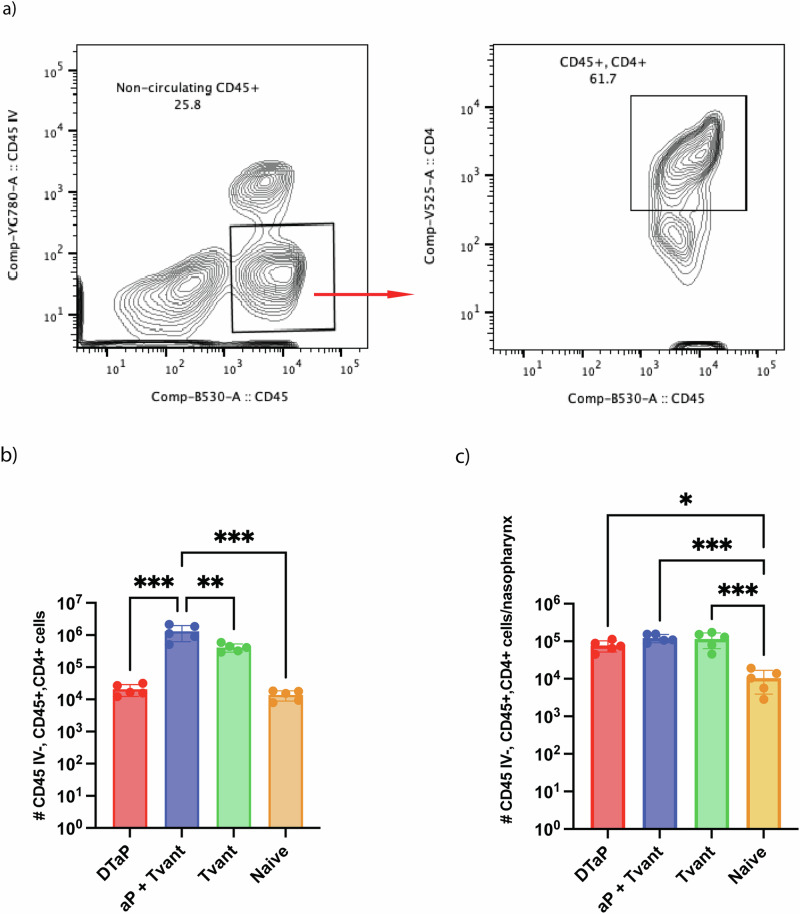
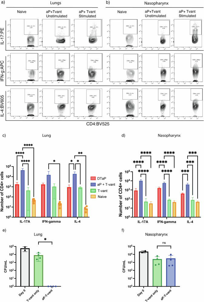
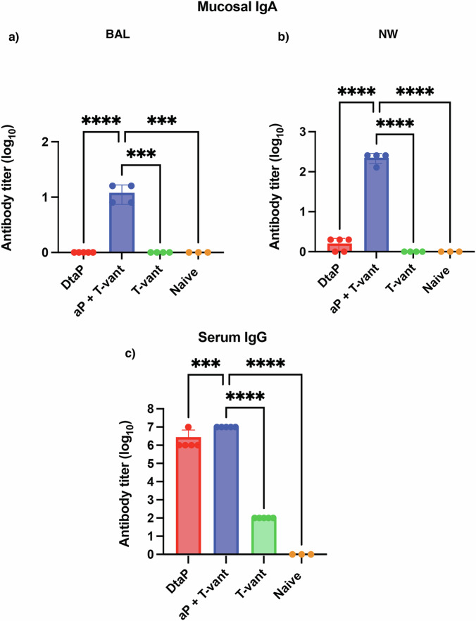
Pertussis is a vaccine-preventable respiratory disease caused by the Gram negative coccobacillus Bordetella pertussis. The licensed acellular pertussis (aP) vaccines protect against disease but do not prevent bacterial colonization and transmission. Here, we developed and tested an intranasal vaccine composed of aP antigens combined with T-vant, a novel adjuvant derived from bacterial outer membrane vesicles, that elicits both mucosal and systemic immune responses. We hypothesized that immunization of mice with aP-T-vant would enhance mucosal immunity and eliminate B. pertussis in the respiratory tract. In contrast to mice immunized intramuscularly with the licensed aP vaccine, intranasal immunization with aP-T-vant eliminated bacteria in both the lung and nasopharynx. Protection was associated with IFN-gamma and IL-17-producing, non-circulating CD4 + T cells in the lung and nasopharynx, and sterilizing immunity in the nasopharynx was dependent on IL-17. Novel mucosal adjuvants, such as T-vant, warrant further investigation to enhance the efficacy of next generation pertussis vaccines.


Conflict of interest statement

J.B.M. and L.A.M. are funded by NIH/NIAID 1R01AI166756-01 and NIH Adjuvant Development Contract 272201800045 C. J.B.M. and L.A.M. are inventors of T-vant and have a patent on the OMV adjuvant platform.

Figures

Fig. 1
Fig. 1. aP-T-vant immunized mice eliminated B. pertussis from the lung and nasopharynx.
a C57Bl/6 mice (n = 6 per group) were immunized three times, 3 weeks apart. Three weeks after the last immunization, mice were challenged intranasally with 1 × 10 < 6 > CFU of Bordetella pertussis strain D420. After infection, lung and nasopharynx were harvested and serial dilutions of tissue homogenates were plated on days 0, 7, 14 and 21. b Mice immunized with aP-T-vant eliminated bacteria from the lung by 14 days post-infection and from the nasopharynx (c) by 21 days post-infection. Limit of detection was 30 CFU per tissue. Data shown is the cumulative data from two independent experiments. ***p < 0.0005, ****p < 0.0001 by 2-way ANOVA of log10 transformed data comparing aP-T-vant to all other groups using a Tukey post-test.
Fig. 2
Fig. 2. aP-T-vant immunization increases CD4 + T cells in the lung and nasopharynx.
PE-Cy7 labeled CD45 antibody was injected in the retro-orbital venous sinus (CD45 IV) of mice (n = 5 per group) 3 min prior to euthanasia. Lung and nasopharynx were harvested, and single cell suspensions were prepared and stained with FITC:CD45 and BV525:CD4 antibodies to identify non-circulating CD4 + T cells. Representative flow cytometry plots are shown for cells gated on CD45 IV- CD4 + . a Non-circulating CD45 + CD4+ cells in (b) lung and (c) nasopharynx. *p < 0.05, **p < 0.005, ***p < 0.0005 by Ordinary one-way ANOVA followed by Tukey post-test.
Fig. 3
Fig. 3. aP-T-vant immunization elicits Th1, Th2 and Th17 producing CD4 + T cells in the lung and nasopharynx.
a Single cell suspensions were prepared from (a) lung and (b) nasopharynx tissues of mice (n = 5 per group) and stimulated in vitro with pooled aP antigens. Intracellular staining for IFN-γ (APC), IL-4 (BV605), and IL-17 (PE) was performed and cells were gated on CD45IV-CD45 + CD4+ by flow cytometry. Representative flow plots are shown for stimulated naive, unstimulated and stimulated aP-T-vant. Increased numbers of cytokine-producing CD4 + T cells were observed in the (c) lung and d nasopharynx of ap-T-vant immunized mice compared to other groups. *p < 0.05, **p < 0.005, ****p < 0.0001 by 2-way ANOVA followed by Tukey’s post-test. e IL-17 is dispensable for bacterial clearance from the lung but is required for clearance in the (f) nasopharynx *p < 0.05 by Mann-Whitney test.
Fig. 4
Fig. 4. T-vant adjuvanted aP elicits mucosal IgA and serum IgG.
Bronchoalveolar lavage (BAL), nasal wash (NW) and sera were obtained 3 weeks post final immunization (n = 3–5 per group). aP-specific reciprocal endpoint titers in the (a) BAL, (b) nasal wash, and (c) sera were measured by ELISA. Ordinary one-way ANOVA followed by Tukey post-test. ***p < 0.0005, ****p < 0.0001.

References

    1. Ryan, M. et al. Distinct T-cell subtypes induced with whole cell and acellular pertussis vaccines in children. Immunology93, 1 (1998). - PMC - PubMed
    1. Warfel, J. M., Zimmerman, L. I. & Merkel, T. J. Comparison of three whole-cell pertussis vaccines in the baboon model of pertussis. Clin. Vacc. Immunol.23, 47–54 (2016). - PMC - PubMed
    1. Wilk, M. M. et al. Immunization with whole cell but not acellular pertussis vaccines primes CD4 TRM cells that sustain protective immunity against nasal colonization with Bordetella pertussis. Emerg. Microbes Infect.8, 169–185 (2019). - PMC - PubMed
    1. Mills, K. H. G., Barnard, A., Watkins, J. & Redhead, K. Cell-mediated immunity to Bordetella pertussis: role of Th1 cells in bacterial clearance in a murine respiratory infection model. Infect. Immun.61, 399–410 (1993). - PMC - PubMed
    1. Warfel, J. M. & Merkel, T. J. Bordetella pertussis infection induces a mucosal IL-17 response and long-lived Th17 and Th1 immune memory cells in nonhuman primates. Mucosal Immunol.6, 787–796 (2013). - PubMed

LinkOut - more resources