. 2024 Oct 15;14(1):24125.
doi: 10.1038/s41598-024-74285-9.

An automatic tracking method to measure the mandibula movement during real time MRI

Affiliations

An automatic tracking method to measure the mandibula movement during real time MRI

Jérémy Mouchoux et al. Sci Rep. .

Abstract

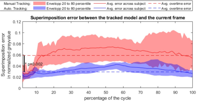
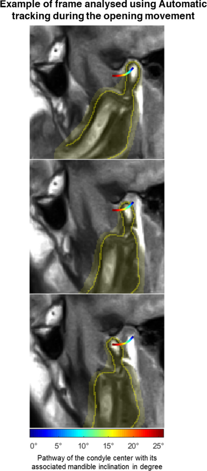
Mandibular movement is complex and individual due to variations in the temporomandibular joint (TMJ). Consequently, patient-centered dentistry should incorporate patients' specific anatomy and condylar function in treatment planning. Real-time magnetic resonance imaging (rt-MRI) visualizes relevant structures and tracks mandibular movement. However, current assessments rely on qualitative observations or time-consuming manual tracking, lacking reliability. This study developed an automatic tracking algorithm for mandibular movement in rt-MRI using least mean square registration (LMS) and compared it to manual tracking (MT) during mouth opening. Ten participants with skeletal class I underwent rt-MRI (10 frames/s). The same operator tracked the condylar pathway for the two methods, setting 2000 landmarks (2 landmarks x100 frames x10 participants) for MT and 210 landmarks (3 landmarks x7 frames x10 participants) for LMS. Time required, superimposition error, and the distance between tracked condylar pathways were compared between methods. LMS tracking was 76% faster and showed significantly better superimposition (0.0289 ± 0.0058) than MT (0.059 ± 0.0145) (p = 0.002). During one-third of the movement, the pathways tracked by both methods were more than 1 mm and 1° apart. These findings highlight the benefits of automatic condylar movement tracking in rt-MRI, laying the groundwork for more objective and quantitative observation of TMJ function.

Keywords: Condyle; LMS algorithm; Temporomandibular joint; Tracking; rtMRI.


Conflict of interest statement

The authors declare no competing interests.

Figures

Fig. 1
Fig. 1
Comparison of the time required for the operator to track the mandible using both methods.
Fig. 2
Fig. 2
Comparison of the error of superimposition obtained with both methods. The automatic tracking is represented in blue while the manual tracking is in red. Its evolution frame per frame averaged across every scan is represented as a plain line and an envelop of lighter color represent the 20 to 80 percentile of the population. Eventually, the averaged error of superimposition across frame and subject is represented as a broken line.
Fig. 3
Fig. 3
Differences between the results of the two tracking methods when assessing the path of the superior condyle, the center of the condyle, the angle to the original inclination, and the path of the ICR.
Fig. 4
Fig. 4
Movement of the Jaw according to the automatic tracking. Three frames (0%, 70% 100%) of the opening movement are displayed. The area of the jaw is highlighted in yellow. The contour of the jaw predicted by the tracking is displayed as yellow dots. The path of the center of the condyle is colored according to the inclination of the jaw.
Fig. 5
Fig. 5
Screenshot of the GUI once the rater is finished. Manual landmarks are not displayed for visibility. They share positions 1 and 3 with automatic tracking. The areas are drawn by the rater and represent the pixels used for the registration. The contours are also drawn by the user but are only used to determine the center of the condyle. Due to the inclination of the acquisition plane, the Processus coronoideus mandibulae is not visible.
Fig. 6
Fig. 6
Grey-values of the pixels within the area drawn by the rater for the mandibula for the current frame in black and for the first frame in red. Horizontal pixels (with vertical tangents) are discarded to reduce computing power. [Matlab, Version R2019a, https://www.mathworks.com/].
Fig. 7
Fig. 7
3D shape extracted from the grey-values for the current frame in black and for the first frame in red This 3D shape of the first frame will be used as a model for the automatic tracking to compute the optimal superposition. [Matlab, Version R2019a, https://www.mathworks.com/].

References

    1. Quast, A. et al. Predictability of maxillary positioning: a 3D comparison of virtual and conventional orthognathic surgery planning. Head Face Med.17, 27 (2021). - PMC - PubMed
    1. Shah, P. & Chong, B. S. 3D imaging, 3D printing and 3D virtual planning in endodontics. Clin. Oral Invest.22, 641–654 (2018). - PubMed
    1. Kernen, F. et al. A review of virtual planning software for guided implant surgery - data import and visualization, drill guide design and manufacturing. BMC Oral Health. 20, 251 (2020). - PMC - PubMed
    1. González-Sánchez, B. et al. Temporomandibular joint dysfunctions: a systematic review of treatment approaches. J. Clin. Med.12 (2023). - PMC - PubMed
    1. Onoda, N., Mikami, T., Saito, N. & Kobayashi, T. Evaluation of oral health-related quality of life in patients with temporomandibular disorders. Cranio - J. Craniomandib.41, 486–493 (2023). - PubMed

LinkOut - more resources