. 2024 Oct 15;14(1):24185.
doi: 10.1038/s41598-024-75127-4.

Examining transfer of TERT to mitochondria under oxidative stress

Affiliations

Examining transfer of TERT to mitochondria under oxidative stress

Dmitrii Burkatovskii et al. Sci Rep. .

Abstract

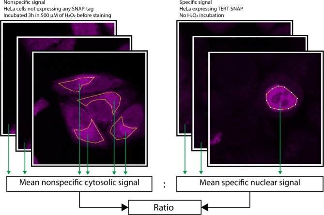
The primary role of telomerase is the lengthening of telomeres. Nonetheless, emerging evidence highlights additional functions of telomerase outside of the nucleus. Specifically, its catalytic subunit, TERT (Telomerase Reverse Transcriptase), is detected in the cytosol and mitochondria. Several studies have suggested an elevation in TERT concentration within mitochondria in response to oxidative stress. However, the origin of this mitochondrial TERT, whether transported from the nucleus or synthesized de novo, remains uncertain. In this study, we investigate the redistribution of TERT, labeled with a SNAP-tag, in response to oxidative stress using laser scanning fluorescence microscopy. Our findings reveal that, under our experimental conditions, there is no discernible transport of TERT from the nucleus to the mitochondria due to oxidative stress.

Keywords: Mitochondria; Oxidative stress; Protein transport; SNAP-tag; TERT.


Conflict of interest statement

The authors declare no competing interests.

Figures

Fig. 1
Fig. 1
TERT-SNAP transport from nucleus to mitochondria under oxidative stress. (A) Experimental concept: TERT-SNAP is initially confined to the cell nucleus labeled with the SNAP-specific dye (depicted as a red star). Following incubation with 500 µM H2O2, two possible scenarios are depicted: left, indicating mature TERT-SNAP transport, resulting in stained mitochondria; right, suggesting the absence of transport, with non-stained mitochondria. (B) Experimental procedure: 1) Staining with SNAP-tag dye. 2) Washing. 3) Initial imaging. 4) Addition of H2O2. 5) Subsequent imaging. (C) Results. Cells are stained with SNAP-SiR (shown as magenta pseudocolor), and H2O2 is introduced to the medium at a concentration of 500 µM. Cell images captured before H2O2 addition (top) and 3 hours after incubation with H2O2 (bottom) illustrate the absence of TERT-SNAP transport within 3 hours.
Fig. 2
Fig. 2
TERT-SNAP synthesis into mitochondria under oxidative stress. (A) Experimental Concept: Before oxidative stress, TERT-SNAP is confined to the cell nucleus labeled with the first SNAP-tag specific dye (indicated as a blue star). The remaining SNAP-tag is blocked with SNAP-Block. Following incubation with 500 µM H2O2, two potential outcomes are depicted: left, illustrating mature TERT-SNAP transport, leading to mitochondria containing TERT-SNAP stained with the first dye; right, representing new TERT-SNAP synthesis, with mitochondria showing no mature TERT-SNAP stained with the first dye. Instead, the second dye will stain the newly synthesized TERT-SNAP within mitochondria. (B) Experimental Procedure: 1) Staining with the first dye. 2) Staining with MitoTracker. 3) Washing. 4) Initial imaging. 5) Application of SNAP-Block. 6) Washing. 7) Addition of H2O2. 8) Staining with the second dye after incubation. 9) Washing. 10) Subsequent imaging. (C) Results: Cells are stained with SNAP-505 (shown as cyan pseudocolor), and MitoTracker Red DND-99 (shown as yellow pseudocolor), while the remaining SNAP-tag is blocked with SNAP-Block. H2O2 is introduced to the medium to a concentration of 500 µM. After 3 hours of incubation, cells are stained with SNAP-SiR (shown as magenta pseudocolor). Consistent with previous experiments, TERT-SNAP stained before H2O2 addition does not appear in mitochondria. The second staining exhibits low specificity, as cells, whether stained or not with SNAP-505 (successfully transfected and not transfected), display similar signal levels from the second dye, SNAP-SiR.
Fig. 3
Fig. 3
Influence of H2O2 incubation on SNAP-Tag dye specificity. The images feature untransfected cells from two distinct dishes with untransfected HeLa cells, each stained with SNAP-SiR (shown as magenta pseudocolor) immediately before imaging. The left images feature cells untreated with H2O2, while the right images depict cells incubated for 3 hours in 500 µM H2O2 before staining and imaging. The H2O2-incubated cells exhibit high nonspecific staining.
Fig. 4
Fig. 4
Nonspecific fluorescence due to H2O2 incubation evaluation. SNAP-SiR signals (shown as magenta pseudocolor) were derived from cells with nonspecifically stained cytosols (left, not transfected, incubated with H2O2) and specifically stained nuclei (right, transfected, not incubated with H2O2). Mean cytosolic and nuclear signals were found, and the ratio between them was calculated.

References

    1. Chan, S. R. W. L. & Blackburn, E. H. Telomeres and telomerase. Philos. Trans. R Soc. Lond. B Biol. Sci. 359, 109–121 (2004). - PMC - PubMed
    1. Zheng, Q., Huang, J. & Wang, G. Mitochondria, telomeres and telomerase subunits. Front. Cell. Dev. Biol. 7, 274 (2019). - PMC - PubMed
    1. Gazzaniga, F. S. & Blackburn, E. H. An antiapoptotic role for telomerase RNA in human immune cells independent of telomere integrity or telomerase enzymatic activity. Blood. 124, 3675–3684 (2014). - PMC - PubMed
    1. Eitan, E. et al. Expression of functional alternative telomerase RNA component gene in mouse brain and in motor neurons cells protects from oxidative stress. Oncotarget. 7, 78297–78309 (2016). - PMC - PubMed
    1. Liu, H., Yang, Y., Ge, Y., Liu, J. & Zhao, Y. TERC promotes cellular inflammatory response independent of telomerase. Nucleic Acids Res. 47, 8084–8095 (2019). - PMC - PubMed

LinkOut - more resources