

Eggplant latent viroid is located in the chloroplasts and nuclei of eggplant infected cells
- PMID: 39407314
- PMCID: PMC11476940
- DOI: 10.1186/s12985-024-02530-8
Eggplant latent viroid is located in the chloroplasts and nuclei of eggplant infected cells
Abstract
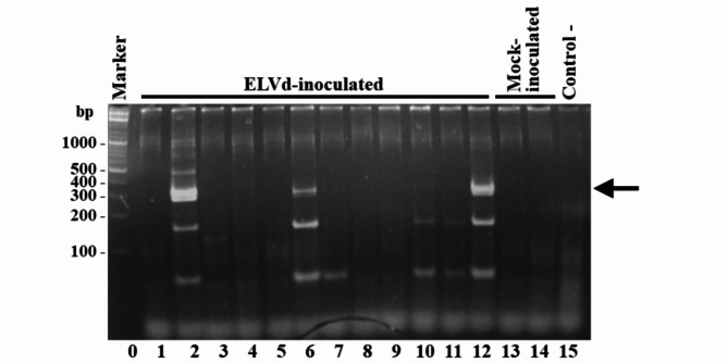
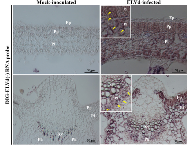
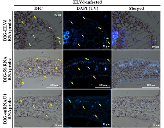
Viroids that belong to genera Avsunviroid and Pelamovirod (family Avsunviroidae) replicate and accumulate in the chloroplasts of infected cells. In this report, we confirmed by RNA in situ hybridization using digoxigenin-UTP-labelled riboprobes that the positive strands of eggplant latent viroid (ELVd), the only member of genus Elaviroid within the family Avsunviroidae, also accumulate in the chloroplasts of infected cells. However, comparison of ELVd in situ hybridization signals with those from bona fide chloroplastic and nuclear non-coding RNAs, such as chloroplast 5S rRNA and U1 small nuclear RNA, supports the notion that this viroid is also present in the nuclei of infected cells. These results suggest that the subcellular localization of viroids within the family Avsunviroidae may be more complex than previously assumed with dynamic presence in several compartments during the infectious cycle.
Keywords: Avsunviroidae; Elaviroid; Non-coding RNA; Subcellular localization; Viroid.
© 2024. The Author(s).
Conflict of interest statement
The authors declare no competing interests.
Figures
References
-
- Adkar-Purushothama CR, Perreault JP. Current overview on viroid–host interactions [Internet]. Wiley Interdiscip. Rev. RNA. Blackwell Publishing Ltd; 2020 [cited 2020 Apr 1]. p. e1570. https://pubmed.ncbi.nlm.nih.gov/31642206/ - PubMed
-
- Navarro B, Flores R, Di Serio F. Advances in Viroid-Host Interactions. Annu Rev Virol [Internet]. 2021 [cited 2021 Oct 23];8:305–25. https://pubmed.ncbi.nlm.nih.gov/34255541/ - PubMed
-
- Ortolá B, Daròs J-A, Viroids. Non-Coding Circular RNAs Able to Autonomously Replicate and Infect Higher Plants. Biology (Basel) [Internet]. 2023;12:172. https://www.mdpi.com/2079-7737/12/2/172 - PMC - PubMed
-
- Itaya A, Folimonov A, Matsuda Y, Nelson RS, Ding B. Potato spindle tuber viroid as inducer of RNA silencing in infected tomato. Mol Plant Microbe Interact [Internet]. 2002/01/05. 2001;14:1332–4. http://www.ncbi.nlm.nih.gov/pubmed/11763132 - PubMed
-
- Papaefthimiou I, Hamilton AJ, Denti MA, Baulcombe DC, Tsagris M, Tabler M. Replicating potato spindle tuber viroid RNA is accompanied by short RNA fragments that are characteristic of post-transcriptional gene silencing. Nucleic Acids Res [Internet]. 2001/05/29. 2001;29:2395–400. http://www.ncbi.nlm.nih.gov/pubmed/11376158 - PMC - PubMed
Publication types
MeSH terms
Substances
LinkOut - more resources
Full Text Sources
