. 2024 Oct 15;12(1):202.
doi: 10.1186/s40168-024-01928-4.

An iron-binding protein of entomopathogenic fungus suppresses the proliferation of host symbiotic bacteria

Affiliations

An iron-binding protein of entomopathogenic fungus suppresses the proliferation of host symbiotic bacteria

Juan Li et al. Microbiome. .

Abstract

Background: Entomopathogenic fungal infection-induced dysbiosis of host microbiota offers a window into understanding the complex interactions between pathogenic fungi and host symbionts. Such insights are critical for enhancing the efficacy of mycoinsecticides. However, the utilization of these interactions in pest control remains largely unexplored.

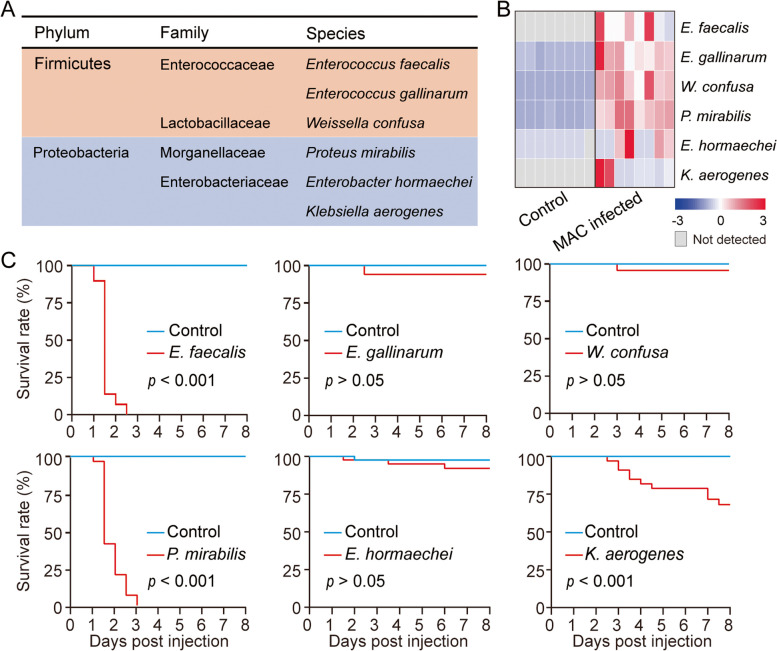
Results: Here, we found that infection by the host-specialist fungus Metarhizium acridum alters the composition of the symbiotic microbiota and increases the dominance of some bacterial symbionts in locusts. Meanwhile, M. acridum also effectively limits the overgrowth of the predominant bacteria. Comparative transcriptomic screening revealed that the fungus upregulates the production of MaCFEM1, an iron-binding protein, in the presence of bacteria. This protein sequesters iron, thereby limiting its availability. Functionally, overexpression of MaCFEM1 in the fungus induces iron deprivation, which significantly suppresses bacterial growth. Conversely, MaCFEM1 knockout relieves the restriction on bacterial iron availability, resulting in iron reallocation. Upon ΔMaCFEM1 infection, some host bacterial symbionts proliferate uncontrollably, turning into opportunistic pathogens and significantly accelerating host death.

Conclusions: This study elucidates the critical role of pathogenic fungal-dominated iron allocation in mediating the shift of host microbes from symbiosis to pathogenicity. It also highlights a unique biocontrol strategy that jointly exploits pathogenic fungi and bacterial symbionts to increase host mortality. Video Abstract.


Conflict of interest statement

The authors declare that they have no competing interests.

Figures

Fig. 1
Fig. 1
M. acridum (MAC) infection causes dysbiosis of microbiota in locusts. A and B Quantifications of fungal (A) and total bacterial (B) loads in the hemolymph of locusts (n = 6, 10 individuals per replicate). Kruskal–Wallis test for multiple comparisons. Values are mean ± standard deviation (SD). Locusts injected with 0.1% Tween 80 serve as the control. C Quantifications of bacterial load in the hemolymph of axenic and non-axenic locusts after MAC injections (n = 4, 10 individuals per replicate). D Survival of axenic and non-axenic locusts after MAC injections (n ≥ 62). Differences between axenic and non-axenic groups are analyzed using the Log-rank test. E Alpha diversity metrics (Chao1 and Shannon index) of bacterial communities in gut of locusts at amplicon sequencing variants (ASV)-level. Differences between control and MAC-infected groups were analyzed using the Mann–Whitney U test, with significant differences denoted as **p < 0.01, and ***p < 0.001. F Principal coordinate analysis (PCoA) at the ASV-level showing the overall differences in gut microbial communities between control and MAC-infected locusts. G Alterations in microbial communities at the family level in the gut of control and MAC-infected locusts. H Bacterial abundance at the family level in hemolymph and gut of locusts after MAC infection
Fig. 2
Fig. 2
Transformation from symbiotic bacteria to opportunistic pathogens results in the death of locusts. A Culturable bacterial isolates derived from hemolymph samples of locusts at 96 h post-infection (hpi) with MAC. B Quantification of relative bacterial levels in locusts’ hemolymph at 96 hpi with MAC using specific 16S rRNA primers (n = 8; 5 locusts per replicate). The heatmap illustrates log2 fold-change values. C Survival of locusts (n = 30) after injection of 5 × 105 colony-forming units (CFUs) of bacterial isolates into the hemocoel. Locusts injected with H2O serve as controls. Differences between the control and bacteria-injected groups are analyzed using the Log-rank test
Fig. 3
Fig. 3
MaCFEM1 is crucial for the colonization of MAC in the presence of symbiotic bacteria. A Experimental procedure for analyzing MAC gene expression in axenic and non-axenic locusts. B Volcano plot showing the overall gene expression pattern of MAC in axenic and non-axenic locusts. C GO enrichment analysis of differentially expressed genes (DEGs), displaying only GO terms with a p value < 0.05. CC, MF, and BP represent cellular components, molecular functions, and biological processes, respectively. D The Venn diagram shows the shared and unique DEGs induced by different bacterial species under fungal-bacterial co-culture conditions. E Gene expression patterns of the candidate DEGs under co-culture conditions. The heat map signal indicates log2 fold-change values of co-cultured groups relative to the control group. MAC cultured alone in ¼SDAY liquid medium serves as the control. Treatments with adjusted p value < 0.05 are denoted by *. F Fungal biomass of WT and mutant strains when co-cultured with E. faecalis, P. mirabilis, S. aureus, and E. coli in ¼SDAY liquid medium, respectively (n = 4). Values are presented as mean ± SD., and differences between WT and mutant strains were analyzed using Student’s t-test, with significant differences denoted as *p < 0.05, **p < 0.01, ***p < 0.001. MaPR1 C, MaCWG, and MaGLUS are gene names for subtilisin-like serine protease PR1C, antigenic cell wall galactomannoprotein, and alpha-glucosidase, respectively
Fig. 4
Fig. 4
MaCFEM1 is an iron-binding protein participating in iron uptake and competition. A Phylogenetic tree illustrates the relationship among MaCFEM1 orthologues in different Metarhizium species. M. acridum and M. album are host-specialists, while M. robertsii, M. brunneum, and M. anisopliae are host-generalists. M. majus and M. guizhouense are transitional species. The phylogenetic tree was conducted using maximum likelihood method. B Gene expression profile of MaCFEM1 under normal and iron-deficient conditions (n = 7). FZ is the abbreviation for Ferrozine, an iron chelator. C Growth of WT and ∆MaCFEM1 under iron-deficient conditions. Relative fungal growth was calculated by normalizing against the area of fungal colonies of the WT grown on the ¼SDAY agar plates. D In vitro expression and purification of MaCFEM1. E Isothermal titration calorimetry analysis for the interaction between Mcfem1 and ferric iron. In three independent experiments, titrating 100 µM proteins into a solution of 10 µM ferric iron resulted in MaCFEM1-Fe3+ N = 3.03 ± 0.86 sites, Ka = 1.69 × 106 ± 8.329 × 105 M − 1, and ∆H =  − 1.17 × 106 ± 7.935 × 105 J/mol. F Schematic diagram for the structure of GFP-MaCFEM1. G Subcellular localization of GFP-MaCFEM1 in MAC hyphae. Scale bar: 10 µm. H MAC upregulates MaCFEM1 expressions when co-cultured with E. faecalis, P. mirabilis, E. coli, and S. aureus, respectively (n = 6). MAC cultured alone in ¼SDAY liquid medium is set as the control. I Iron content per gram of mycelium (n = 10). J CFU of E. faecalis and P. mirabilis per milliliter of liquid medium under co-culture conditions (n = 5). Values are presented as mean ± SD, One-way ANOVA with Bonferroni's test for multiple comparisons (C and H), Mann–Whitney U test (B, I, and J) for pairwise comparisons. Significant differences are denoted by ***p < 0.001 or by different letters
Fig. 5
Fig. 5
∆MaCFEM1 infection causes uncontrolled proliferation of opportunistic bacteria, accelerating locust death. A Expression of MaCFEM1 under axenic and non-axenic conditions (n = 5, 8 locusts/replicate). B and C Survival rate of non-axenic and axenic locusts following WT and ∆MaCFEM1 injections or topical infections (n ≥ 30). Locusts treated with 0.1% Tween 80 were used as controls. D Quantification of total bacterial and MAC fungal loads in non-axenic and axenic locusts at 96 hpi. E Quantification of E. faecalis and P. mirabilis loads in non-axenic and axenic locusts at 96 hpi (n ≥ 5, 8 locusts/replicate). F Iron content in hemolymph of locusts at 96 hpi with WT and ∆MaCFEM1 (n = 5, 20 individuals/replicate). G Cadavers of MAC-infected locusts were categorized into three types based on body color: red, brown, and black. H Bacterial load in different cadavers (n = 5, 8 cadavers/replicate). I Ratio of different cadavers calculated following WT and ∆MaCFEM1 infections (n ≥ 90). J Expression levels of MaCFEM1 in WT and MaCFEM1 overexpression strain (OE-MaCFEM1) in vivo, and bacterial load at 96 hpi with WT and OE-MaCFEM1 (n = 4, 8 locusts/replicate). K Survival rate of locusts after WT and OE-MaCFEM1 infections (n ≥ 60). Locusts injected with 0.1% Tween 80 serve as the control. A, D, E, I, and J Values represent mean ± SD. Pairwise comparisons were performed using Student’s t-test or Mann–Whitney U test, and significant differences are indicated by **p < 0.01, ***p < 0.001. n.s represents no significant differences. H One-way ANOVA with Bonfferroni’s test for multiple comparisons. Values are mean ± SD, significant differences are denoted by different letters. K Log-rank test for survival dynamics analysis
Fig. 6
Fig. 6
The proposed model demonstrates that disruption of the iron competition-driven microbial equilibrium can trigger symbiotic bacteria to transform into opportunistic pathogens, thereby elevating host mortality. Infection by M. acridum disrupts the composition of the locust microbiota, leading to the dominance of some symbiotic bacteria and prompting their migration from the gut to the hemocoel. Meanwhile, the fungus maintains a delicate balance with these bacteria in the hemocoel by restraining their overgrowth, thereby efficiently exploiting host resources. In the presence of bacteria, M. acridum upregulates the iron-binding protein MaCFEM1, which sequesters iron, thus restricting bacterial access to iron. Knockout of MaCFEM1 removes these fungal-imposed limitations on bacterial iron acquisition, resulting in unregulated bacterial proliferation. Consequently, locusts infected with the ΔMaCFEM1 exhibit high mortality and shortened lifespans due to the uncontrolled proliferation of opportunistic bacteria

Similar articles

Cited by

References

    1. Robinson CM. Enteric viruses exploit the microbiota to promote infection. Curr Opin Virol. 2019;37:58–62. - PMC - PubMed
    1. Jia D, Mao Q, Chen Y, Liu Y, Chen Q, Wu W, Zhang X, Chen H, Li Y, Wei T. Insect symbiotic bacteria harbour viral pathogens for transovarial transmission. Nat Microbiol. 2017;2:17025. - PubMed
    1. Kim EK, Lee KA, Hyeon DY, Kyung M, Jun KY, Seo SH, Hwang D, Kwon Y, Lee WJ. Bacterial nucleoside catabolism controls quorum sensing and commensal-to-pathogen transition in the gut. Cell Host Microbe. 2020;27(3):345–57. - PubMed
    1. Wei G, Lai Y, Wang G, Chen H, Li F, Wang S. Insect pathogenic fungus interacts with the gut microbiota to accelerate mosquito mortality. Proc Natl Acad Sci U S A. 2017;114(23):5994–9. - PMC - PubMed
    1. Narasimhan S, Rajeevan N, Liu L, Zhao YO, Heisig J, Pan JY, Eppler-Epstein R, DePonte K, Fish D, Fikrig E. Gut microbiota of the tick vector modulate colonization of the lyme disease spirochete. Cell Host Microbe. 2014;15(1):58–71. - PMC - PubMed

LinkOut - more resources