

Substantia nigra hyperechogenicity and brain ventricular size as biomarkers of early dementia with Lewy bodies
- PMID: 39407323
- PMCID: PMC11475835
- DOI: 10.1186/s13195-024-01590-w
Substantia nigra hyperechogenicity and brain ventricular size as biomarkers of early dementia with Lewy bodies
Abstract
Background: Diagnosis of dementia with Lewy bodies (DLB) is challenging, especially in the earlier stages of the disease, owing to the clinical overlap with other neurodegenerative diseases such as Alzheimer's (AD) and Parkinson's disease (PD). We aimed to identify the transcranial sonography (TCS) parameters that can help us to detect early DLB patients.
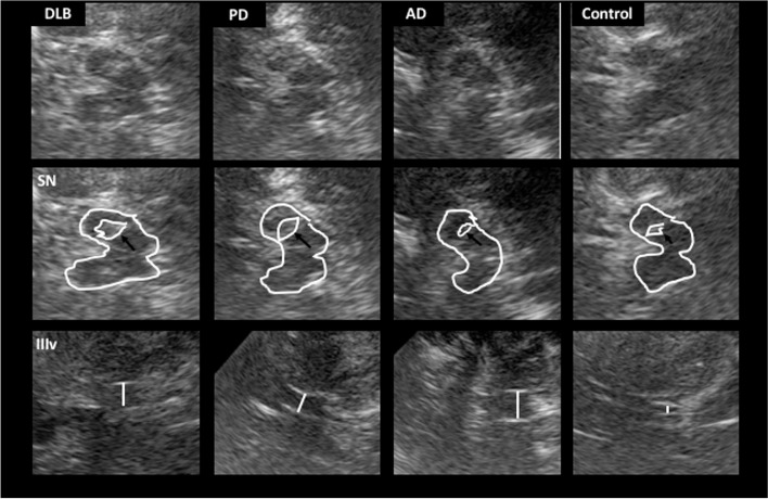
Methods: In this cross-sectional study, we prospectively recruited newly diagnosed DLB patients with less than 3 years from the onset of cognitive symptoms. For comparison purposes, we also included AD and PD patients, with a disease duration of less than 3 years, and a control group. TCS was performed to assess the substantia nigra (SN) echogenicity, the width of the third ventricle, and the frontal horns of the lateral ventricles. Subsequently, TCS images were analyzed with the medical image viewer Horos in order to quantify the intensity of the echogenicity of the SN. Univariate analysis and a logistic regression model were used to identify which variables can predict the diagnosis of DLB.
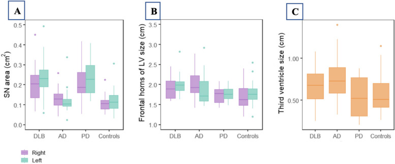
Results: One hundred and seven participants were included (23 DLB, 26 AD, 27 PD and 31 controls). The median age of DLB patients was 75(72-77) years, with a disease duration of 2 years. DLB and PD patients showed higher SN hyperechogenicity rates (72.73% and 81.82%, respectively) and a greater area of the SN compared to AD patients and controls (p < 0.001). DLB and AD patients had wider ventricular systems than the other study groups. The SN hyperechogenicity predicted a diagnosis of DLB with an odds ratio of 22.67 (95%CI 3.98; 129.12, p < 0.001) when compared to AD patients. Unilateral and bilateral widened frontal horns predicted diagnosis of DLB compared to PD with an odds ratio of 9.5 (95%CI 0.97; 92.83, p = 0.053) and 5.7 (95%CI 0.97; 33.6, p = 0.054), respectively.
Conclusions: Echogenicity of the SN and widening of the frontal horns of lateral ventricles can predict the diagnosis of early DLB in this cohort of newly diagnosed patients, when compared to AD and PD patients. Transcranial sonography, a non-invasive tool, could be helpful for the diagnosis of DLB at its earlier stages.
Keywords: Dementia with Lewy bodies; Frontal horns of lateral ventricles; Substantia nigra; Third ventricle width; Transcranial sonography.
© 2024. The Author(s).
Conflict of interest statement
The authors declare no competing interests.
Figures
References
MeSH terms
Substances
LinkOut - more resources
Full Text Sources
Medical
