. 2024 Sep 30;29(19):4654.
doi: 10.3390/molecules29194654.

1,2,4-Oxadiazole Derivatives: Physicochemical Properties, Antileishmanial Potential, Docking and Molecular Dynamic Simulations of Leishmania infantum Target Proteins

Affiliations

1,2,4-Oxadiazole Derivatives: Physicochemical Properties, Antileishmanial Potential, Docking and Molecular Dynamic Simulations of Leishmania infantum Target Proteins

Deyzi C S Barbosa et al. Molecules. .

Abstract

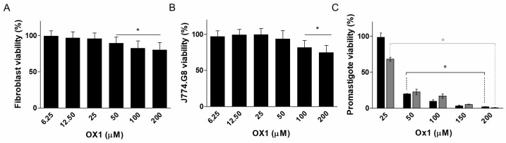
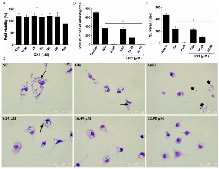
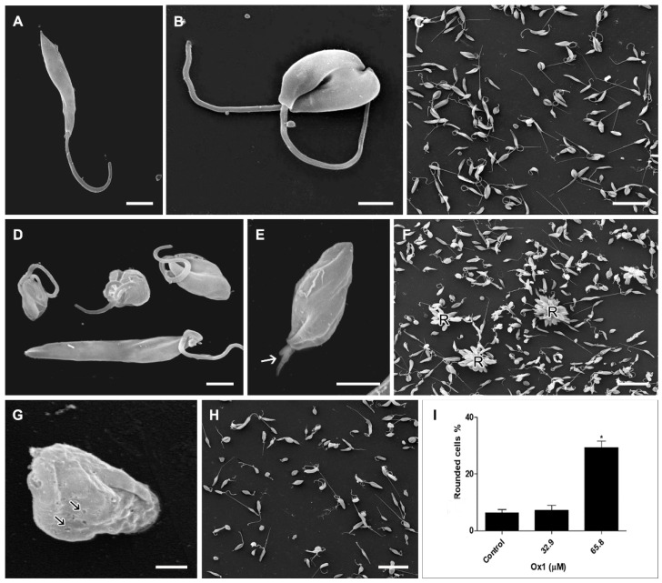
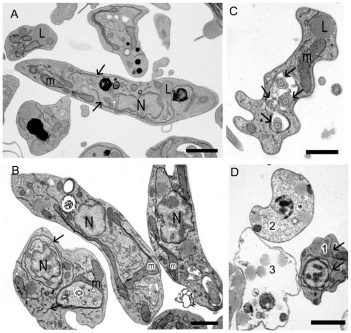
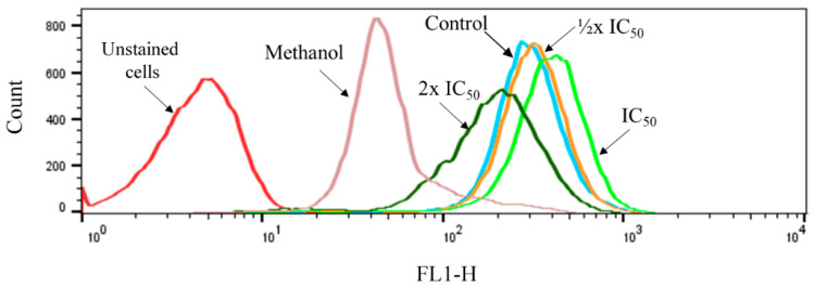
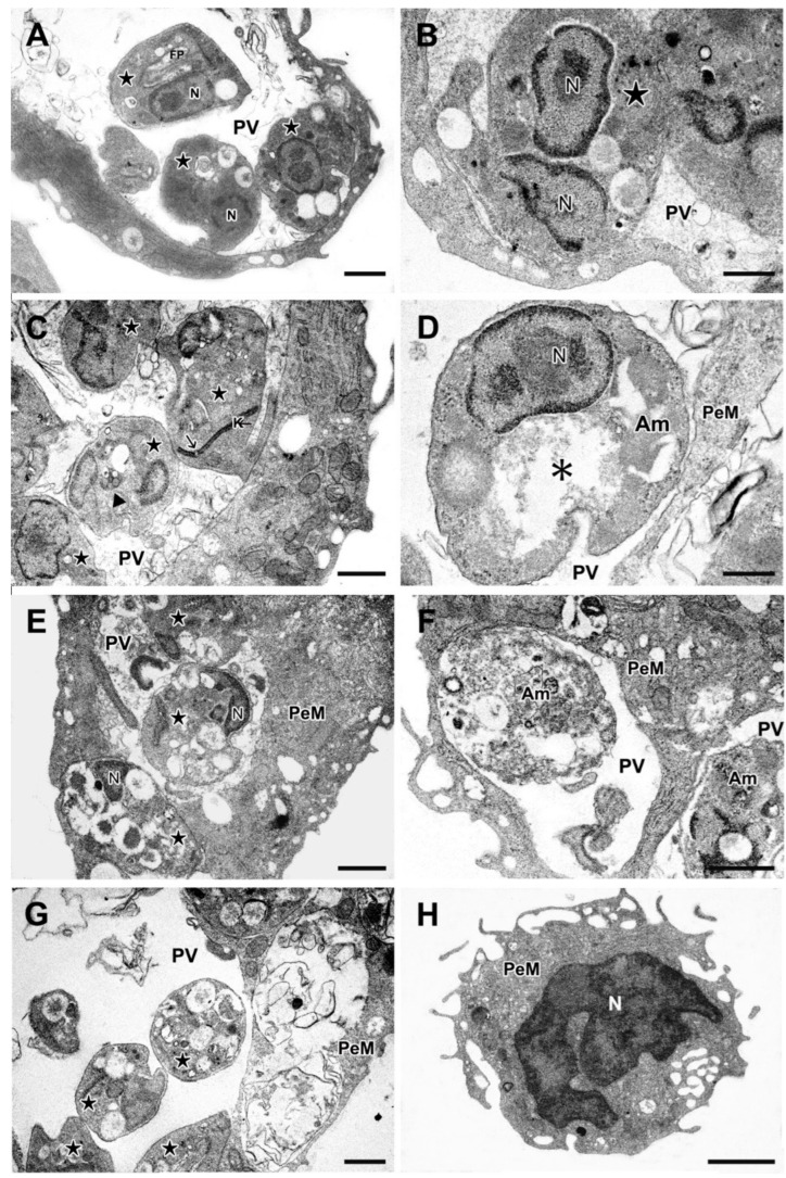
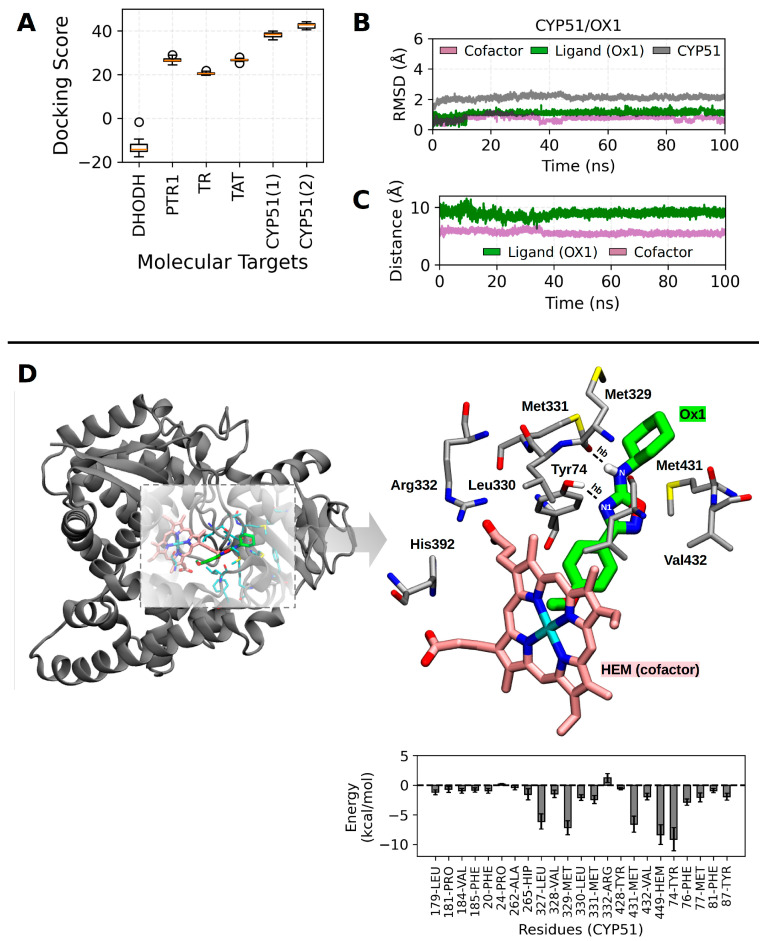
Visceral leishmaniasis (VL), caused by protozoa of the genus Leishmania, remains a significant public health concern due to its potentially lethal nature if untreated. Current chemotherapy options are limited by severe toxicity and drug resistance. Derivatives of 1,2,4-oxadiazole have emerged as promising drug candidates due to their broad biological activity. This study investigated the effects of novel 1,2,4-oxadiazole derivatives (Ox1-Ox7) on Leishmania infantum, the etiological agent of VL. In silico predictions using SwissADME suggest that these compounds have high oral absorption and good bioavailability. Among them, Ox1 showed the most promise, with higher selectivity against promastigotes and lower cytotoxicity towards L929 fibroblasts and J774.G8 macrophages. Ox1 exhibited selectivity indices of 18.7 and 61.7 against L. infantum promastigotes and amastigotes, respectively, compared to peritoneal macrophages. Ultrastructural analyses revealed severe morphological damage in both parasite forms, leading to cell death. Additionally, Ox1 decreased the mitochondrial membrane potential in promastigotes, as shown by flow cytometry. Molecular docking and dynamic simulations indicated a strong affinity of Ox1 for the L. infantum CYP51 enzyme. Overall, Ox1 is a promising and effective compound against L. infantum.

Keywords: 1,2,4-oxadiazole; chemotherapy; molecular docking; molecular dynamic; ultrastructure; visceral leishmaniasis.


Conflict of interest statement

The authors declare no conflicts of interest.

Figures

Scheme 1
Scheme 1
Representative scheme of the synthesis of 1,2,4-oxadiazole derivatives Ox1Ox7.
Figure 1
Figure 1
Representative graphs of the effects of Ox1 on mammalian cells and promastigotes. (A) Activity of Ox1 on the viability of fibroblast (L929) and (B) macrophages (J774.G8) after 48 h of drug treatment. (C) Effects of Ox1 on promastigotes after 24 (black bars) and 48 (gray bars) hours of treatment. Values represent the mean ± standard deviation of three independent experiments in triplicate. * Significant differences at p < 0.05 compared to untreated control.
Figure 2
Figure 2
Effects of Ox1 on the viability of PeMs and intracellular amastigotes of forms of L. infantum. (A) Percentage of viable treated PeMs compared to untreated ones. (B) Total number of amastigotes in 300 infected cells. (C) Survival index (SuI) of amastigotes in PeMs. Glu—glucantime; AmB—Amphotericin B. Each bar represents the mean ± SD of three independent experiments performed in duplicate. * Significant differences at p < 0.05 compared to the untreated control. (D) Representative images of untreated infected-PeMs negative control (NC) or treated with Glu, AmB, or Ox1 at ¼ (8.24 µM), ½ IC50 (16.42 µM), and IC50 (32.98 µM) for promastigote forms. Intracellular amastigotes are indicated by arrows. Note in the culture treated with AmB the presence of cellular debris (thick arrow). Cells treated with IC50 and ½ IC50 of Ox1 or AmB, presenting empty parasitophorous vacuoles (*) can be also observed. Bars 25 µm.
Figure 3
Figure 3
Scanning electron microscopy of control and treated promastigotes of L. infantum. (A) Detail of untreated promastigote showing elongated cell body, smooth plasma membrane, and long flagellum. (B) Untreated dividing promastigotes showing a preserved morphology. (C) Low magnification of untreated culture showing the predominance of elongated and dividing cells. (D) treated culture showing rounded cells with twisted flagellum around the parasite cell body next to an elongated cell. (E) High magnification of treated cell showing rounded cell body, septation, and altered and short flagellum (arrow). (F) Low magnification of culture treated with IC50 Ox1 showing numerous promastigote rosettes (R). (G) Severe injured cells treated with 2× IC50 Ox1. Note the presence of cell membrane perforations (arrow), the loss of characteristic morphology, and the absence of visible flagellum. (H) At low magnification, it is possible to observe the decrease in cell number and the increase in cells presenting altered rounded morphology. (I) Representative graph of the percentage of rounded promastigotes treated with IC50 (32.98 µM) and 2× IC50 (65.96 µM). A total of 500 cells were randomly counted per sample. * Significant differences at p < 0.05 compared to the untreated control. Bars: (A,B,D,E) =2 µm; (C,F,H) =20 µm; (G) =1 µm.
Figure 4
Figure 4
Transmission electron microscopy of control and treated promastigotes. (A) Untreated promastigote showing dispersed endoplasmic reticulum (arrow), preserved nucleus (N), mitochondrion (m), and lipid droplets (L). (B) Promastigote treated with IC50 Ox1 showing altered mitochondrion (asterisk) and increased endoplasmic reticulum (arrow). (C) Promastigote treated with 2× IC50 showing multiple flagella within the flagellar pocket (arrow). (D) Note the presence of varying levels of cellular damage (1–3), increase in endoplasmic reticulum (1), and lipid droplets (2 and 3). Bars: (A,B)—2 µm. (C,D)—1 µm.
Figure 5
Figure 5
Representative histogram of untreated cells (control) and cells treated with half, once, and twice the IC50 of Ox1.
Figure 6
Figure 6
Transmission electron microscopy of the effects of Ox1 on (PeM) infected with amastigote. (A,B) Untreated infected cells displaying several amastigotes (star) inside the parasitophorous vacuole (PV). (B) Detail of dividing amastigote (star) with preserved cellular structure. (C,D) Infected macrophages treated with IC50 Ox1. (C) Amastigotes (stars) presenting cytoplasmic vacuoles surrounded by membrane (arrowhead). Stretched kinetoplast showing partial rupture can be observed (arrow). (D) Amastigote with loss of cytoplasmic content, asterisk. (E,F) Infected PeM treated with 2× IC50 Ox1. (E) Amastigotes (star) presenting numerous membrane-bound vacuoles with or without internal content. Note the presence of high amounts of cellular debris inside the PV. (F) Detail of partially degraded amastigote (Am) inside the PV. (G) Infected macrophages treated with Amphotericin B. Amastigotes were indicated by stars. (H) Non-infected and non-treated macrophage. PV, parasitophorous vacuole; Am, amastigote; PeM, peritoneal macrophages infected with amastigote; FP, flagellar pocket; N, nucleus; K, kinetoplast. Bars: (A,C,E,F) =1µm; (G,H) =2 µm; (B,D) =500 nm.
Figure 7
Figure 7
Summary of the in-silico results. (A) Docking scores obtained with GOLD software; label (1) corresponds to the scores obtained with the Chemscore scoring function, (2) corresponds to the modified version of this scoring function, with iron parameters. (B) RMSD profile during MD simulation. (C) Analysis of the center of mass distance during MD simulation. (D) The CYP51/Ox1 complex is shown on the left and a detailed view of the binding site is shown on the right, showing the hydrophobic contacts and two hydrogen bonds (hb) between CYP51 and Ox1 compound: (i) the hydroxyl group of Tyr74 and the nitrogen atom (N1) of Ox1, and (ii) the oxygen atom of Met329 and hydrogen atom attached to the nitrogen (N) to the Ox1. The interaction energy profile between CYP51 (per residue) and Ox1 is shown below. It is important to note that the Ox1 binding pose does not reflect the interaction analysis entirely since this analysis was carried out considering more than one binding pose. The structure of the CYP51 receptor in the cartoon model is shown in gray, and the carbon atoms of the binding site are in sticks model and colored in gray, whereas the carbon atoms of Ox1 are shown in green. The carbon atoms of the heme propionate are shown in pink. The hydrogen atoms were omitted to improve visualization.

Similar articles

References

    1. Wamai R.G., Kahn J., McGloin J., Ziaggi G. Visceral Leishmaniasis: A Global Overview. J. Glob. Health Sci. 2020;2:e3. doi: 10.35500/jghs.2020.2.e3. - DOI
    1. Van Griensven J., Diro E. Visceral Leishmaniasis. Infect. Dis. Clin. N. Am. 2012;26:309–322. doi: 10.1016/j.idc.2012.03.005. - DOI - PubMed
    1. Soni V., Chandel S., Pandey V., Asati S., Jain P., Jain A., Tekade R.K. Chapter 8—Novel Therapeutic Approaches for the Treatment of Leishmaniasis. In: Tekade R.K., editor. Biomaterials and Bionanotechnology. Academic Press; Cambridge, MA, USA: 2019. pp. 263–300. Advances in Pharmaceutical Product Development and Research.
    1. Mann S., Frasca K., Scherrer S., Henao-Martínez A.F., Newman S., Ramanan P., Suarez J.A. A Review of Leishmaniasis: Current Knowledge and Future Directions. Curr. Trop. Med. Rep. 2021;8:121–132. doi: 10.1007/s40475-021-00232-7. - DOI - PMC - PubMed
    1. WHO Leishmaniasis. [(accessed on 29 June 2024)]. Available online: https://www.who.int/news-room/fact-sheets/detail/leishmaniasis.

MeSH terms

LinkOut - more resources