Review
. 2024 Oct 1;29(19):4672.
doi: 10.3390/molecules29194672.

Nanoparticle Tracking Analysis: An Effective Tool to Characterize Extracellular Vesicles

Affiliations
Review

Nanoparticle Tracking Analysis: An Effective Tool to Characterize Extracellular Vesicles

Gabrielle Kowkabany et al. Molecules. .

Abstract

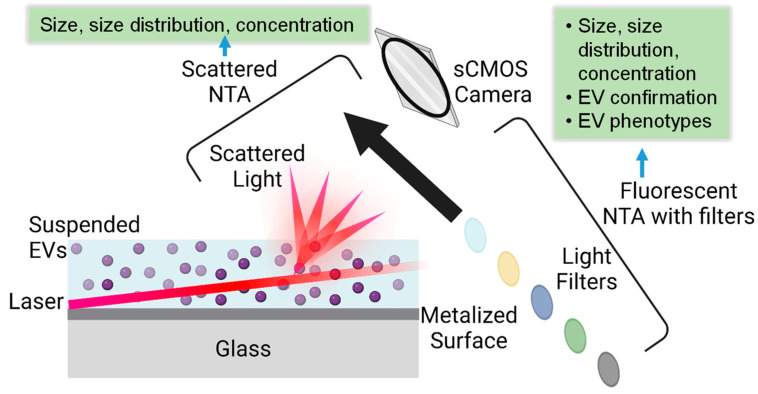
Extracellular vesicles (EVs) are membrane-enclosed particles that have attracted much attention for their potential in disease diagnosis and therapy. However, the clinical translation is limited by the dosing consistency due to their heterogeneity. Among various characterization techniques, nanoparticle tracking analysis (NTA) offers distinct benefits for EV characterization. In this review, we will discuss the NTA technique with a focus on factors affecting the results; then, we will review the two modes of the NTA techniques along with suitable applications in specific areas of EV studies. EVs are typically characterized by their size, size distribution, concentration, protein markers, and RNA cargos. The light-scattering mode of NTA offers accurate size, size distribution, and concentration information in solution, which is useful for comparing EV isolation methods, storage conditions, and EV secretion conditions. In contrast, fluorescent mode of NTA allows differentiating EV subgroups based on specific markers. The success of fluorescence NTA heavily relies on fluorescent tags (e.g., types of dyes and labeling methods). When EVs are labeled with disease-specific markers, fluorescence NTA offers an effective tool for disease detection in biological fluids, such as saliva, blood, and serum. Finally, we will discuss the limitations and future directions of the NTA technique in EV characterization.

Keywords: exosomes; extracellular vesicles; fluorescent labeling; nanoparticle-tracking analysis.


Conflict of interest statement

The authors declare no conflicts of interest.

Figures

Figure 1
Figure 1
Illustration of NTA measurement principle in scattering and fluorescent modes along with obtained EV information, where the various colored circles indicated that different wavelength of emission lights can be collected by the selection of appropriate filter.
Figure 2
Figure 2
Illustration of various labeling strategies of EVs, such as lipophilic dyes for non-specific EV labeling, a fluorescent antibody targeting EV surface markers, or fluorescent protein (FP) fused to either surface proteins or internal cytosolic proteins, and mRNA labeling.
Figure 3
Figure 3
Single EV analysis by a multifluorescence NTA with time-sequential illumination where CD81, CD9, and CD63 markers were labeled by blue, green and red fluorescence, respectively: (a) illustration of the detection process, (b) relative percentage of EVs with and without fluorescent labels, and (c) comparison of detection of EVs with three CD markers by NTA and TIRF. (Copyright © 2021 American Chemical Society).

Similar articles

Cited by

References

    1. van Niel G., D’Angelo G., Raposo G. Shedding light on the cell biology of extracellular vesicles. Nat. Rev. Mol. Cell Biol. 2018;19:213–228. doi: 10.1038/nrm.2017.125. - DOI - PubMed
    1. Gurung S., Perocheau D., Touramanidou L., Baruteau J. The exosome journey: From biogenesis to uptake and intracellular signalling. Cell Commun. Signal. 2021;19:47. doi: 10.1186/s12964-021-00730-1. - DOI - PMC - PubMed
    1. Hessvik N.P., Llorente A. Current knowledge on exosome biogenesis and release. Cell. Mol. Life Sci. 2018;75:193–208. doi: 10.1007/s00018-017-2595-9. - DOI - PMC - PubMed
    1. He C.J., Zheng S., Luo Y., Wang B. Exosome Theranostics: Biology and Translational Medicine. Theranostics. 2018;8:237–255. doi: 10.7150/thno.21945. - DOI - PMC - PubMed
    1. Lobb R.J., Lima L.G., Moller A. Exosomes: Key mediators of metastasis and pre-metastatic niche formation. Semin. Cell Dev. Biol. 2017;67:3–10. doi: 10.1016/j.semcdb.2017.01.004. - DOI - PubMed

LinkOut - more resources