Review
. 2024 Sep 28;13(19):5791.
doi: 10.3390/jcm13195791.

Optical Coherence Tomography in Myocardial Infarction Management: Enhancing Precision in Percutaneous Coronary Intervention

Affiliations
Review

Optical Coherence Tomography in Myocardial Infarction Management: Enhancing Precision in Percutaneous Coronary Intervention

Angela Buonpane et al. J Clin Med. .

Abstract

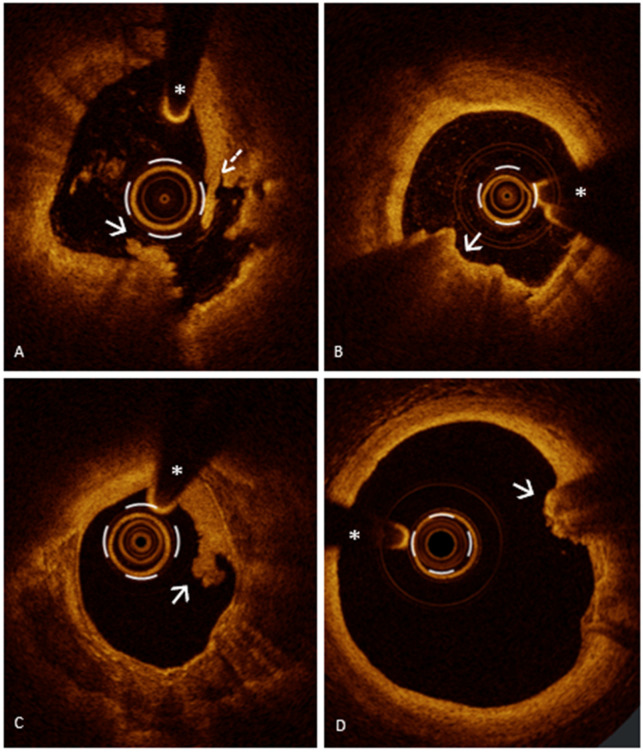
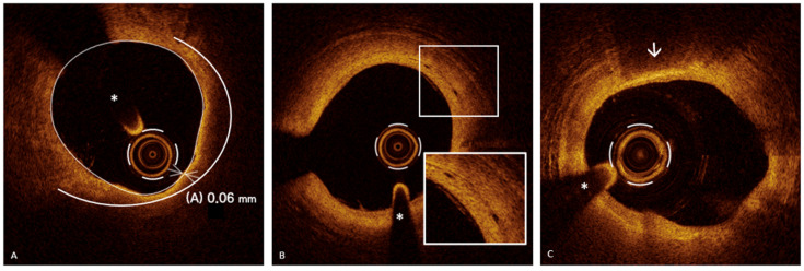
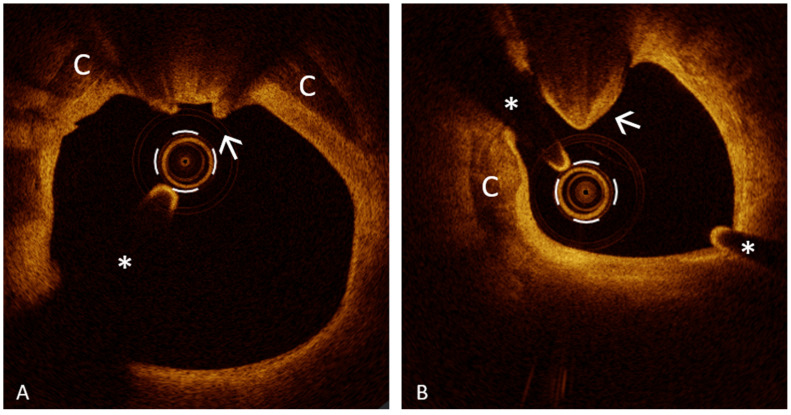
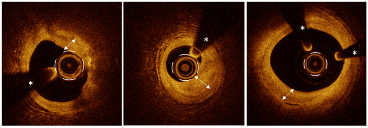
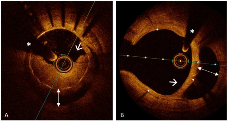
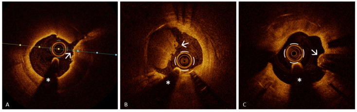
In acute myocardial infarction (AMI), the urgency of coronary revascularization through percutaneous coronary intervention (PCI) is paramount, offering notable advantages over pharmacologic treatment. However, the persistent risk of adverse events, including recurrent AMI and heart failure post-revascularization, underscores the necessity for enhanced strategies in managing coronary artery disease. Traditional angiography, while widely employed, presents significant limitations by providing only two-dimensional representations of complex three-dimensional vascular structures, hampering the accurate assessment of plaque characteristics and stenosis severity. Intravascular imaging, specifically optical coherence tomography (OCT), significantly addresses these limitations with superior spatial resolution compared to intravascular ultrasound (IVUS). Within the context of AMI, OCT serves dual purposes: as a diagnostic tool to accurately identify culprit lesions in ambiguous cases and as a guide for optimizing PCI procedures. Its capacity to differentiate between various mechanisms of acute coronary syndrome, such as plaque rupture and spontaneous coronary dissection, enhances its diagnostic potential. Furthermore, OCT facilitates precise lesion preparation, optimal stent sizing, and confirms stent deployment efficacy. Recent meta-analyses indicate that OCT-guided PCI markedly improves safety and efficacy in revascularization, subsequently decreasing the risks of mortality and complications. This review emphasizes the critical role of OCT in refining patient-specific therapeutic approaches, aligning with the principles of precision medicine to enhance clinical outcomes for individuals experiencing AMI.

Keywords: acute myocardial infarction; mechanisms of acute coronary syndrome; optical coherence tomography; precision medicine.


Conflict of interest statement

The authors declare no conflicts of interest.

Figures

Figure 1
Figure 1
Mechanisms of myocardial infarction: PR and PE. Panel (A). PR: rupture of the fibrous cap resulting in a large vessel wall cavity with the exposure of highly thrombotic necrotic core to the blood flow; evidence of mixed thrombus (arrow) and white thrombus (dashed arrow) at both sites of the cavity. Panel (B,C); definite OCT-erosion: presence of thrombus overlying an intact plaque without discontinuity of the fibrous cap. Panel (B): red thrombus at 6–7 o’clock (white arrow) with posterior shadowing, precluding interpretation of the underlying tissue. Panel (C): white thrombus at 2–3 o’clock (white arrow) with low backscattering and without posterior shadowing allowing for the interpretation of the underlying plaque phenotype, a very calcified plaque with diffuse calcification (calcium arc of about 270°). Panel (D); probable OCT-erosion: presence of red thrombus (high backscattering and posterior shadowing) with attenuation of the underlying plaque (white arrow) without atherosclerotic features proximal or distal to its site. The asterisk is indicating the guide wire artifact.
Figure 2
Figure 2
Plaque vulnerability features. Panel (A); thin cap fibroatheroma (TCFA): lipid-rich plaque with a lipid arc > 180° (white semicircle) covered by a fibrous cap < 65 µm at its thinnest point (A). Panel (B); microvessels: round- to oval-shaped structures with black content (“small black holes”) and a diameter of 50–300 μm (magnification in insert). Panel (C); macrophages: highly intense backscattering “bright spot”, casting a dark shadow which is sharply bordered laterally. A thin layer of macrophages is observed at 12 o’clock (white arrow). The asterisk is indicating the guide wire artifact.
Figure 3
Figure 3
Calcified nodule (CN): calcified lesion protruding into the coronary artery lumen. Panel (A): Eruptive CN with fibrous cap discontinuity and overlying mixed thrombus (white arrow) in the context of severely calcified atherosclerotic disease. Spotty calcium is indicated with (C). Panel (B); noneruptive CN: Calcified lesion protruding into the vessel lumen without evidence of fibrous cap discontinuity and overlying thrombus (white arrow). Spotty calcium is indicated with (C). The asterisk is indicating the guide wire artifact.
Figure 4
Figure 4
Healed plaque: layered structure with an “onion-like” appearance, featuring one or more layers with intense, heterogeneous signals, layers of different optical densities (double-headed arrow), and a distinct demarcation from the underlying tissue. The asterisk is indicating the guide wire artifact.
Figure 5
Figure 5
Stent malapposition: separation of at least one stent strut from the intimal surface of the coronary artery wall with a distance between the strut’s surface to the luminal surface greater than the strut thickness. Panel (A): in-stent thrombosis with a mixed thrombus (white arrow) associated with the major stent malapposition (double-head arrow) of the previous implanted stent. Panel (B): major stent malapposition (double-head arrow) with neointimal hyperplasia (white arrow) embedding the stent struts (resulting in a “dual” lumen appearance). The asterisk is indicating the guide wire artifact.
Figure 6
Figure 6
Neoatherosclerosis: the presence of at least one component of a mature atherosclerotic plaque, such as lipid-rich tissue or calcification, within the neointima. Panel (A): PR (with arrow) with overlying microthrombi in neoatherosclerosis with unstable plaque characteristics (lipid-laden neointima with a thin fibrous cap). Panel (B): definite OCT-erosion with mixed thrombus (with arrow) in fibrocalcific neoatherosclerosis. Panel (C): eruptive CN with irregular surface (with arrow) in neoatherosclerosis with diffuse calcification. The asterisk is indicating the guide wire artifact.

Similar articles

Cited by

References

    1. Bottardi A., Prado G.F.A., Lunardi M., Fezzi S., Pesarini G., Tavella D., Scarsini R., Ribichini F. Clinical Updates in Coronary Artery Disease: A Comprehensive Review. J. Clin. Med. 2024;13:4600. doi: 10.3390/jcm13164600. - DOI - PMC - PubMed
    1. Giubilato S., Lucà F., Abrignani M.G., Gatto L., Rao C.M., Ingianni N., Amico F., Rossini R., Caretta G., Cornara S., et al. Management of Residual Risk in Chronic Coronary Syndromes. Clinical Pathways for a Quality-Based Secondary Prevention. J. Clin. Med. 2023;12:5989. doi: 10.3390/jcm12185989. - DOI - PMC - PubMed
    1. Paradossi U., Taglieri N., Massarelli G., Palmieri C., De Caterina A.R., Bruno A.G., Taddei A., Nardi E., Ghetti G., Palmerini T., et al. Female Gender and Mortality in ST-Segment-Elevation Myocardial Infarction Treated with Primary PCI. J. Cardiovasc. Med. 2022;23:234–241. doi: 10.2459/JCM.0000000000001300. - DOI - PubMed
    1. Paradossi U., De Caterina A.R., Trimarchi G., Pizzino F., Bastiani L., Dossi F., Raccis M., Bianchi G., Palmieri C., de Gregorio C., et al. The Enigma of the ‘Smoker’s Paradox’: Results from a Single-Center Registry of Patients with STEMI Undergoing Primary Percutaneous Coronary Intervention. Cardiovasc. Revasc. Med. 2024. in Press . - DOI - PubMed
    1. Marchi F., Pylypiv N., Parlanti A., Storti S., Gaggini M., Paradossi U., Berti S., Vassalle C. Systemic Immune-Inflammation Index and Systemic Inflammatory Response Index as Predictors of Mortality in ST-Elevation Myocardial Infarction. J. Clin. Med. 2024;13:1256. doi: 10.3390/jcm13051256. - DOI - PMC - PubMed

LinkOut - more resources