

Electrocardiogram Features of Left Ventricular Excessive Trabeculation with Preserved Cardiac Function in Light of Cardiac Magnetic Resonance and Genetics
- PMID: 39407966
- PMCID: PMC11477278
- DOI: 10.3390/jcm13195906
Electrocardiogram Features of Left Ventricular Excessive Trabeculation with Preserved Cardiac Function in Light of Cardiac Magnetic Resonance and Genetics
Abstract
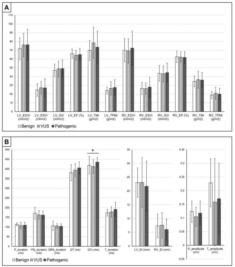
Background and Objectives: Although left ventricular excessive trabeculation (LVET) can cause heart failure, arrhythmia and thromboembolism, limited literature is available on the ECG characteristics of primary LVET with preserved left ventricular function (EF). We aimed to compare the ECG characteristics and cardiac MR (CMR) parameters of LVET individuals with preserved left ventricular EF to a control (C) group, to identify sex-specific differences, and to compare the genetic subgroups of LVET with each other and with a C population. Methods: In our study, we selected 69 LVET individuals (EF > 50%) without any comorbidities and compared them to 69 sex- and age-matched control subjects (42% females in both groups, p = 1.000; mean age LVET-vs-C: 38 ± 14 vs. 38 ± 14 years p = 0.814). We analyzed the pattern and notable parameters of the 12-lead ECG recordings. We determined the volumetric and functional parameters, as well as the muscle mass values of the left and right ventricles (LV, RV) based on the CMR recordings. Based on the genotype, three subgroups were established: pathogenic, variant of uncertain significance and benign. Results: In the LVET group, we found normal but elevated volumetric and muscle mass values and a decreased LV_EF, wider QRS, prolonged QTc, higher RV Sokolow index values and lower T wave amplitude compared to the C. When comparing MR and ECG parameters between genetic subgroups, only the QTc showed a significant difference. Over one-third of the LVET population had arrhythmic episodes and a positive family history. Conclusions: The subclinical morphological and ECG changes and the clinical background of the LVET group indicate the need for follow-up of this population, even with preserved EF.
Keywords: ECG; cardiac magnetic resonance imaging; cardiogenetics; left ventricular excessive trabeculation; noncomapction.
Conflict of interest statement
The authors declare no conflicts of interest. The funders had no role in the design of the study; in the collection, analyses, or interpretation of data; in the writing of the manuscript; or in the decision to publish the results.
Figures
References
-
- Petersen S.E., Jensen B., Aung N., Friedrich M.G., McMahon C.J., Mohiddin S.A., Pignatelli R.H., Ricci F., Anderson R.H., Bluemke D.A. Excessive Trabeculation of the Left Ventricle: JACC: Cardiovascular Imaging Expert Panel Paper. JACC Cardiovasc. Imaging. 2023;16:408–425. doi: 10.1016/j.jcmg.2022.12.026. - DOI - PMC - PubMed
Grants and funding
LinkOut - more resources
Full Text Sources
