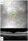
Microperimetry Sensitivity Correlates to Structural Macular Changes in Adolescents with Achromatopsia Unlike Other Visual Function Tests
- PMID: 39408028
- PMCID: PMC11478056
- DOI: 10.3390/jcm13195968
Microperimetry Sensitivity Correlates to Structural Macular Changes in Adolescents with Achromatopsia Unlike Other Visual Function Tests
Abstract
Objectives: Achromatopsia (ACHM) is a rare autosomal, recessively inherited disease that is characterized by cone dysfunction, for which several gene therapies are currently on trial. The aim of this study was to find correlations between the morphological macular changes identified using optical coherence tomography (OCT) and some visual functional parameters. Visual acuity (VA), contrast sensitivity (CS), and macular sensitivity obtained by means of microperimetry were assessed. Methods: Adolescents with ACHM underwent macular microperimetry (S-MAIA device) in mesopic condition, macular OCT, best corrected visual acuity (BCVA), low luminance visual acuity (LLVA), near vision acuity (NVA), and CS measurement. Results: Eight patients (15 eyes) with ACHM were analyzed. The mean age was 17 ± 2.7 years, and genetic variants involved the CNGA3 gene (37.5%) and CNGB3 gene (62.5%). OCT staging significantly correlated with microperimetry sensitivity parameters, namely the sensitivity of the central foveal point (p = 0.0286) and of the first and second perifoveal rings (p = 0.0008 and p = 0.0014, respectively). No correlations were found between OCT staging and VA measurements, nor with CS value. Conclusions: Among the extensive evaluated visual function tests, only microperimetry sensitivity showed a correlation with morphological macular changes identified at OCT. Microperimetry sensitivity may thus represent a useful visual function tool in natural ACHM history studies considering the upcoming research on gene therapies for the treatment of ACHM.
Keywords: achromatopsia; best corrected visual acuity; cone dystrophy; contrast sensitivity; low luminance visual acuity; microperimetry; optical coherence tomography; retinal inherited disease.
Conflict of interest statement
The authors declare no conflicts of interest.
Figures

Similar articles
-
Phenotypic characteristics of Danish patients with achromatopsia.Acta Ophthalmol. 2024 Sep;102(6):e893-e905. doi: 10.1111/aos.16656. Epub 2024 Feb 13. Acta Ophthalmol. 2024. PMID: 38348755
-
Long-Term Investigation of Retinal Function in Patients with Achromatopsia.Invest Ophthalmol Vis Sci. 2020 Sep 1;61(11):38. doi: 10.1167/iovs.61.11.38. Invest Ophthalmol Vis Sci. 2020. PMID: 32960951 Free PMC article.
-
MORPHOFUNCTIONAL EVALUATION OF MACULAR-FOVEAL CAPILLARIES: A Comparative Optical Coherence Tomography Angiography and Microperimetry Study.Retina. 2020 Jul;40(7):1279-1285. doi: 10.1097/IAE.0000000000002616. Retina. 2020. PMID: 31274711
-
[Correlation of retinal sensitivity, visual acuity and central macular thickness in different types of diabetic macular edema].Zhonghua Yan Ke Za Zhi. 2013 Dec;49(12):1081-8. Zhonghua Yan Ke Za Zhi. 2013. PMID: 24499694 Chinese.
-
Achromatopsia: Genetics and Gene Therapy.Mol Diagn Ther. 2022 Jan;26(1):51-59. doi: 10.1007/s40291-021-00565-z. Epub 2021 Dec 3. Mol Diagn Ther. 2022. PMID: 34860352 Free PMC article. Review.
References
-
- Sharpe L.T., Stockman A., Jagle H., Nathans J. Opsin genes, cone photopigments, color vision, and color blindness. In: Gegenfurtner K., Sharpe L.T., editors. Color Vision: From Genes to Perception. Cambridge University Press; Cambridge, UK: 1999. pp. 3–52.
-
- Kohl S., Zobor D., Chiang W., Weisschuh N., Staller J., Menendez I.G., Chang S., Beck S.C., Garrido M.G., Sothilingam V., et al. Mutations in the unfolded protein response regulator ATF6 cause the cone dysfunction disorder achromatopsia. Nat. Genet. 2015;47:757–765. doi: 10.1038/ng.3319. - DOI - PMC - PubMed
-
- Thiadens A.A.H.J., Somervuo V., van den Born L.I., Roosing S., van Schooneveld M.J., Kuijpers R.W.A.M., van Moll-Ramirez N., Cremers F.P.M., Hoyng C.B., Klaver C.C.W. Progressive Loss of Cones in Achromatopsia: An Imaging Study Using Spectral-Domain Optical Coherence Tomography. Investig. Ophthalmol. Vis. Sci. 2010;51:5952–5957. doi: 10.1167/iovs.10-5680. - DOI - PubMed
LinkOut - more resources
Full Text Sources

