. 2024 Sep 30;16(19):3323.
doi: 10.3390/nu16193323.

Isatidis Folium Represses Dextran Sulfate Sodium-Induced Colitis and Suppresses the Inflammatory Response by Inhibiting Inflammasome Activation

Affiliations

Isatidis Folium Represses Dextran Sulfate Sodium-Induced Colitis and Suppresses the Inflammatory Response by Inhibiting Inflammasome Activation

You Chul Chung et al. Nutrients. .

Abstract

Background/objectives: Isatidis Folium (IF) has been used in traditional medicine for various ailments, and recent research highlights its anti-inflammatory, antiviral, and detoxifying properties. This study investigated the anti-inflammatory effects of a hydroethanolic extract of IF (EIF) on inflammasomes and colitis.

Methods: Dextran sulfate sodium (DSS)-induced colitis model C57BL/6 mice were treated with DSS, mesalamine, or EIF (200 mg/kg). Parameters such as daily disease activity index (DAI), spleen weight, colon length, and histopathology were evaluated. Intestinal fibrosis, mucin, and tight junction proteins were assessed using Masson's trichrome, periodic acid-Schiff, and immunohistochemistry staining. RAW264.7 and J774a.1 macrophages were treated with EIF and lipopolysaccharide, with cell viability assessed via the cell counting kit-8 assay, nitric oxide (NO) production with Griess reagent, and cytokine levels with the enzyme-linked immunosorbent assay. NF-κB inhibition was analyzed using the luciferase assay, and phytochemical analysis was performed using UPLC-MS/MS.

Results: EIF mitigated weight loss, reduced DAI scores, prevented colon shortening, and attenuated mucosal damage, fibrosis, and goblet cell loss while enhancing the tight junction protein occludin. The anti-inflammatory effects of EIF in RAW264.7 cells included reduced NO production, pro-inflammatory cytokines, and NF-κB activity, along with inhibition of NLRP3 inflammasome responses in J774a.1 cells. The key constituents identified were tryptanthrin, indigo, and indirubin.

Conclusions: Animal studies demonstrated the efficacy of EIF in alleviating colitis, suggesting its potential for treating inflammatory diseases.

Keywords: Isatidis Folium; anti-inflammatory agents; caspase-1; inflammasome; macrophages; ulcerative colitis.


Conflict of interest statement

The authors declare no conflicts of interest.

Figures

Figure 1
Figure 1
Therapeutic effects of the hydroethanolic extract of Isatidis Folium (EIF) on dextran sulfate sodium (DSS)-induced colitis in mice (n = 8). (A) The body weight change profile showed that EIF treatment mitigated weight loss. (B) The Disease Activity Index (DAI) indicated reduced colitis severity with EIF. Data are presented as means ± standard errors; ** p < 0.01, and **** p < 0.0001 versus the negative control (N.C.) group (two-way ANOVA with Dunnett’s comparison as the post hoc test). (C) Colon length comparison demonstrated less shortening in EIF-treated mice, and the spleen index indicated reduced spleen enlargement. Data are presented as means ± standard errors; * p < 0.05 and *** p < 0.001 versus the N.C. group (two-tailed Student’s t-test). (D) Hematoxylin and eosin (H&E)-stained colonic sections revealed reduced inflammation and tissue damage in EIF-treated mice. Data are presented as means ± standard errors; *** p < 0.001 versus N.C. group (one-way ANOVA with Dunnett’s comparison as the post hoc test).
Figure 2
Figure 2
The histopathological changes in DSS-induced ulcerative colitis were evaluated. (A) Masson’s trichrome (MT) staining revealed collagen deposition indicative of fibrosis in colonic sections. (B) Periodic acid–Schiff (PAS) staining showed goblet cell preservation and mucin production. Immunohistochemistry (IHC) staining demonstrated the expression of tight junction proteins, with (C) ZO-1 and (D) occludin. The data were analyzed using one-way ANOVA with Dunnett’s post hoc test, presented as means ± standard errors. Statistical significance was marked as * p < 0.05, and *** p < 0.001 versus the N.C. group. n = 8 (A,B) and n = 6 (C,D). ns., not significant.
Figure 3
Figure 3
Effects of EIF on inflammasome component gene expression in DSS-induced ulcerative colitis (n = 4). RT-qPCR analysis of (A) NLRP3, (B) caspase-1, (C) ASC, and (D) IL-1β in the intestines of DSS-induced colitis mice. Data are presented as the means ± standard errors from three independent experiments. Statistical analysis was performed using one-way ANOVA followed by Dunnett’s post hoc test. Statistical significance compared to the N.C. group is indicated as * p < 0.05, ** p < 0.01, and *** p < 0.001.
Figure 4
Figure 4
Modulatory effects of the hydroethanolic extract of Isatidis Folium (EIF) on LPS-induced inflammatory responses in RAW264.7 macrophages. (A) Cell viability was determined using the Cell Counting Kit-8 (CCK-8) assay, showing that EIF maintained cell viability in LPS-treated RAW264.7 cells within a non-cytotoxic range. (B) Nitric oxide (NO) production was measured by the Griess reagent assay. (C) Tumor necrosis factor-alpha (TNF-α), (D) interleukin-6 (IL-6), and (E) interleukin-1 beta (IL-1β) levels were significantly lowered by EIF treatment, assessed via ELISA. (F) NF-κB activity was suppressed by EIF, as indicated by the luciferase reporter assay. Dexamethasone (DEX) was used as a positive control. Data are presented as the means ± standard errors from three independent experiments. Statistical analysis was performed using one-way ANOVA followed by Dunnett’s post hoc test. Statistical significance compared to the N.C. group is indicated as * p < 0.05, ** p < 0.01, and *** p < 0.001. N.D., not detected.
Figure 5
Figure 5
Expression of inflammasome component proteins (NLRP3, ASC, and caspase-1) and IL-1β in J774a.1 cells treated with EIF. (A) Western blot analysis showed levels of NLRP3, ASC, caspase-1, IL-1β, and β-actin. Quantification of protein levels revealed (B) reduced NLRP3, (C) decreased caspase-1, and (D) lower IL-1β expression in EIF-treated cells compared to those in the LPS/ATP-treated group. MCC950, an NLRP3 inflammasome inhibitor, was used as a positive control. Data are presented as the means ± standard errors from three independent experiments. Statistical analysis was performed using one-way ANOVA followed by Dunnett’s post hoc test. Statistical significance compared to the LPS/ATP-treated group is indicated as * p < 0.05, ** p < 0.01, and *** p < 0.001.
Figure 6
Figure 6
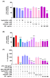
Effect of EIF constituents on J774a.1 macrophages. (A) Cell viability was assessed in cells treated only with EIF and its components. In LPS/ATP-treated cells, EIF constituents were tested for their effect on (B) cell viability and (C) IL-1β expression. MCC950, an NLRP3 inflammasome inhibitor, was used as the positive control. Data are presented as the means ± standard errors from three independent experiments. Statistical analysis was performed using one-way ANOVA followed by Dunnett’s post hoc test. Statistical significance compared to the LPS/ATP-treated group is indicated as * p < 0.05, ** p < 0.01, and *** p < 0.001.
Figure 7
Figure 7
Expression of inflammasome component proteins and IL-1β in J774a.1 macrophage cells treated with EIF constituents. In LPS/ATP-treated J774a.1 cells, EIF constituents were tested for their effect on protein expression. (A) Western blot analysis and protein levels of (B) NLRP3, (C) caspase-1, and (D) IL-1β. MCC950, an NLRP3 inflammasome inhibitor, was used as a positive control. Data are presented as the means ± standard errors from three independent experiments. Statistical analysis was performed using one-way ANOVA followed by Dunnett’s post hoc test. Statistical significance compared to the LPS/ATP-treated group is indicated as * p < 0.05, ** p < 0.01, and *** p < 0.001.
Figure 8
Figure 8
Effect of EIF constituents on bone marrow-derived macrophages (BMDMs). (A) Cell viability was assessed in cells treated only with EIF and its components. In LPS/ATP-treated BMDMs, EIF constituents were tested for their effects on (B) cell viability and (C) IL-1β expression. MCC950, an NLRP3 inflammasome inhibitor, was used as a positive control. Data are presented as the means ± standard errors from three independent experiments. Statistical analysis was performed using one-way ANOVA followed by Dunnett’s post hoc test. Statistical significance compared to the LPS/ATP-treated group is indicated as * p < 0.05, ** p < 0.01, and *** p < 0.001. N.D., not detected.

Similar articles

Cited by

References

    1. Baumgart D.C., Sandborn W.J. Crohn’s disease. Lancet. 2012;380:1590–1605. doi: 10.1016/S0140-6736(12)60026-9. - DOI - PubMed
    1. Podolsky D.K. Inflammatory bowel disease. N. Engl. J. Med. 2002;347:417–429. doi: 10.1056/NEJMra020831. - DOI - PubMed
    1. Abraham C., Medzhitov R. Interactions between the host innate immune system and microbes in inflammatory bowel disease. Gastroenterology. 2011;140:1729–1737. doi: 10.1053/j.gastro.2011.02.012. - DOI - PMC - PubMed
    1. Kaser A., Zeissig S., Blumberg R.S. Inflammatory bowel disease. Annu. Rev. Immunol. 2010;28:573–621. doi: 10.1146/annurev-immunol-030409-101225. - DOI - PMC - PubMed
    1. Sartor R.B. Microbial influences in inflammatory bowel diseases. Gastroenterology. 2008;134:577–594. doi: 10.1053/j.gastro.2007.11.059. - DOI - PubMed

LinkOut - more resources