. 2024 Sep 24;25(19):10268.
doi: 10.3390/ijms251910268.

Three-Dimensional-Bioprinted Non-Small Cell Lung Cancer Models in a Mouse Phantom for Radiotherapy Research

Affiliations

Three-Dimensional-Bioprinted Non-Small Cell Lung Cancer Models in a Mouse Phantom for Radiotherapy Research

Yikun Mei et al. Int J Mol Sci. .

Abstract

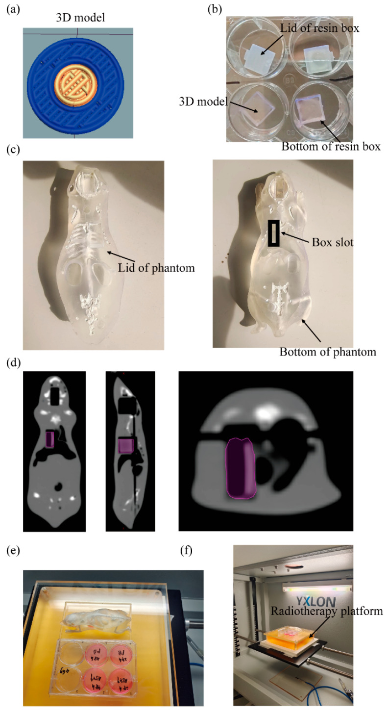
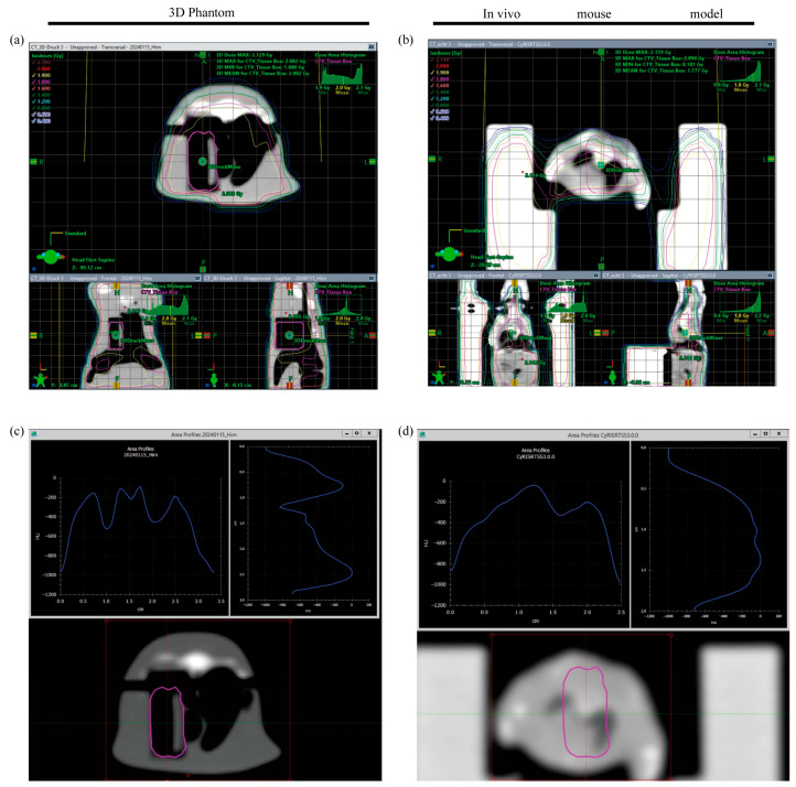
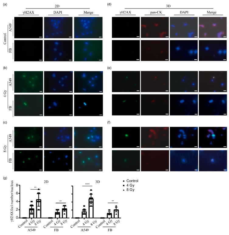
Lung cancer continues to have one of the highest morbidity and mortality rates of any cancer. Although radiochemotherapy, in combination with immunotherapy, has significantly improved overall survival, new treatment options are urgently needed. However, preclinical radiotherapy testing is often performed in animal models, which has several drawbacks, including species-specific differences and ethical concerns. To replace animal models, this study used a micro-extrusion bioprinting approach to generate a three-dimensional (3D) human lung cancer model consisting of lung tumor cells embedded in human primary lung fibroblasts for radiotherapy research. The models were placed in a mouse phantom, i.e., a 3D-printed mouse model made of materials that mimic the X-ray radiation attenuation rates found in mice. In radiotherapy experiments, the model demonstrated a selective cytotoxic effect of X-rays on tumor cells, consistent with findings in 2D cells. Furthermore, the analysis of metabolic activity, cell death, apoptosis, and DNA damage-induced γH2AX foci formation revealed different results in the 3D model inside the phantom compared to those observed in irradiated models without phantom and 2D cells. The proposed setup of the bioprinted 3D lung model inside the mouse phantom provides a physiologically relevant model system to study radiation effects.

Keywords: 3D bioprinting; lung cancer; model system; mouse phantom; radiation therapy.


Conflict of interest statement

The authors declare no conflicts of interest.

Figures

Figure 1
Figure 1
Design of the 3D-printed human lung cancer model and arrangement of the radiotherapy equipment. (a) Schematic representation of the 3D model. (b) Three-dimensional models in resin boxes. (c) Upper and lower parts of the mouse phantom. (d) Coronal, sagittal, and axial CT images of the mouse phantom. The purple area highlights the thoracic slot where the resin box containing the 3D model can be placed in the mouse lung. (e) Experimental setup of 2D cell culture plates and the mouse phantom for radiation. (f) The entire internal irradiation area of the radiotherapy device.
Figure 2
Figure 2
Dose distribution (marked by colored isodose curves) as well as mean irradiation doses were comparable between the 3D phantom (a) and the in vivo mouse model (b). Investigation of density histograms showed differences according to anatomical localization (e.g., lower Houndsfield units in lung tissue) (c,d). Clinical target volume (pink contour) was delineated by hand on a CT scan of the 3D phantom and manually transferred to a CT scan of the in vivo mouse model [16].
Figure 3
Figure 3
Cell viability and metabolic activity of 2D cells and 3D models after irradiation. The metabolic activity of 2D lung fibroblasts (FB) (a) and A549 cells (b) was measured by a tetrazolium hydroxide salt (XTT) assay at the indicated time points after irradiation with 4 or 8 Gy, respectively. The control groups were kept under the same conditions as the two experimental groups, except that they were not irradiated. Data from three independent experiments are presented as mean ± standard deviation, **** p < 0.0001. (ce) The viability of 2D cells was evaluated for the non-irradiated control (c), 4 Gy irradiation group (d), and 8 Gy irradiation group (e), at 24 h post-radiotherapy using calcein-AM and ethidium homodimer-1 staining. Under fluorescence microscopy, viable cells appeared green, while dead cells appeared red. Scale bar: 200 µm. (f) The metabolic activity of the 3D lung cancer models was measured using an XTT assay at 24 h, 48 h, and 5 days after irradiation, and the data were plotted for radiation doses of 4 Gy and 8 Gy. The control group was maintained under the same conditions as the two experimental groups except that it was not irradiated. Data from three independent experiments are presented as mean ± standard deviation, **** p < 0.0001. (g) Cell viability staining of the 3D lung cancer models was conducted at 24 h post-radiotherapy using calcein-AM and ethidium homodimer-1 staining. The cancer part containing A549 cells is marked with a red circle. Scale bar: 500 µm.
Figure 4
Figure 4
γH2AX immunofluorescence staining of irradiated 2D cells and 3D models. (ac) Immunostaining images of A549 cells and lung fibroblasts (FB) from the unirradiated control (a), 4 Gy irradiation group (b), and 8 Gy irradiation group (c). Cells were fixed 24 h after irradiation. γH2AX staining (green channel) indicates DNA double-strand breaks. DAPI was used for nuclear counterstaining (blue channel). Scale bar: 20 µm. (df) Immunohistochemical staining images of 3D lung cancer models from the non-irradiated control group (d), 4 Gy irradiation group (e), and 8 Gy irradiation group (f). The models were fixed, dehydrated, and paraffin-embedded 24 h after irradiation, followed by sectioning at 16 µm thickness. The sections were then subjected to immunostaining. γH2AX staining (green channel) indicates DNA double-strand breaks. The samples were also stained with antibodies against pan-cytokeratin (pan-CK) to confirm their identity as epithelial cells (red channel). DAPI was used for nuclear counterstaining (blue channel). Scale bar: 20 µm. (g) After treatment with different radiation doses, the average number of γH2AX foci was determined in the nuclei of randomly selected A549 cells and fibroblasts in 2D, as well as in regions of A549 cells and fibroblasts within 3D model sections. Data from three independent experiments are presented as mean ± standard deviation, ** p < 0.01, **** p < 0.0001.
Figure 5
Figure 5
Cell viability and metabolic activity of 3D models after irradiation without the mouse phantom. (a) The metabolic activity of the 3D lung cancer models placed in a 12-well plate was measured using an XTT assay 24 h after irradiation with 4 or 8 Gy, respectively. The control group was maintained under the same conditions as the two experimental groups except that it was not irradiated. Data from three independent experiments are expressed as the mean ± standard deviation, **** p < 0.0001. (b) Cell viability staining of the 3D lung cancer models placed in a 12-well plate 24 h after irradiation using calcein-AM and ethidium homodimer-1 staining. Under fluorescence microscopy, viable cells appeared green, while dead cells appeared red. The cancer part containing A549 cells is marked with a red circle. Scale bar: 500 µm.

Similar articles

Cited by

References

    1. Siegel R.L., Miller K.D., Fuchs H.E., Jemal A. Cancer Statistics, 2021. CA-Cancer J. Clin. 2021;71:7–33. doi: 10.3322/caac.21654. - DOI - PubMed
    1. Oliver A.L. Lung Cancer: Epidemiology and Screening. Surg. Clin. N. Am. 2022;102:335–344. doi: 10.1016/j.suc.2021.12.001. - DOI - PubMed
    1. Brown S., Banfill K., Aznar M.C., Whitehurst P., Faivre Finn C. The evolving role of radiotherapy in non-small cell lung cancer. Br. J. Radiol. 2019;92:20190524. doi: 10.1259/bjr.20190524. - DOI - PMC - PubMed
    1. Meagan M., Nasser H. Advances in systemic therapy for non-small cell lung cancer. BMJ. 2021;375:n2363. - PubMed
    1. Vinod S.K., Hau E. Radiotherapy treatment for lung cancer: Current status and future directions. Respirology. 2020;25((Suppl. S2)):61–71. doi: 10.1111/resp.13870. - DOI - PubMed

LinkOut - more resources