. 2024 Sep 26;25(19):10382.
doi: 10.3390/ijms251910382.

Enhanced Photodynamic Therapy Efficacy through Solid Lipid Nanoparticle of Purpurin-18-N-Propylimide Methyl Ester for Cancer Treatment

Affiliations

Enhanced Photodynamic Therapy Efficacy through Solid Lipid Nanoparticle of Purpurin-18-N-Propylimide Methyl Ester for Cancer Treatment

Sooho Yeo et al. Int J Mol Sci. .

Abstract

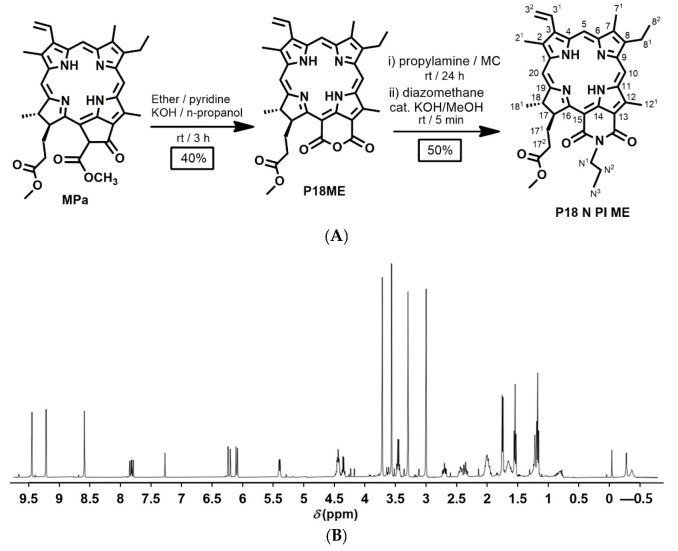
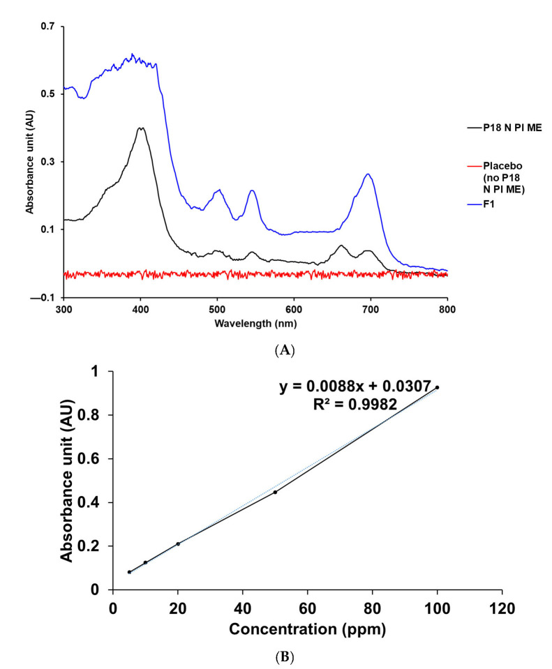
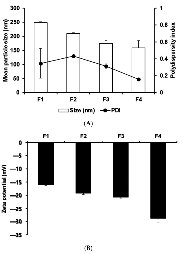
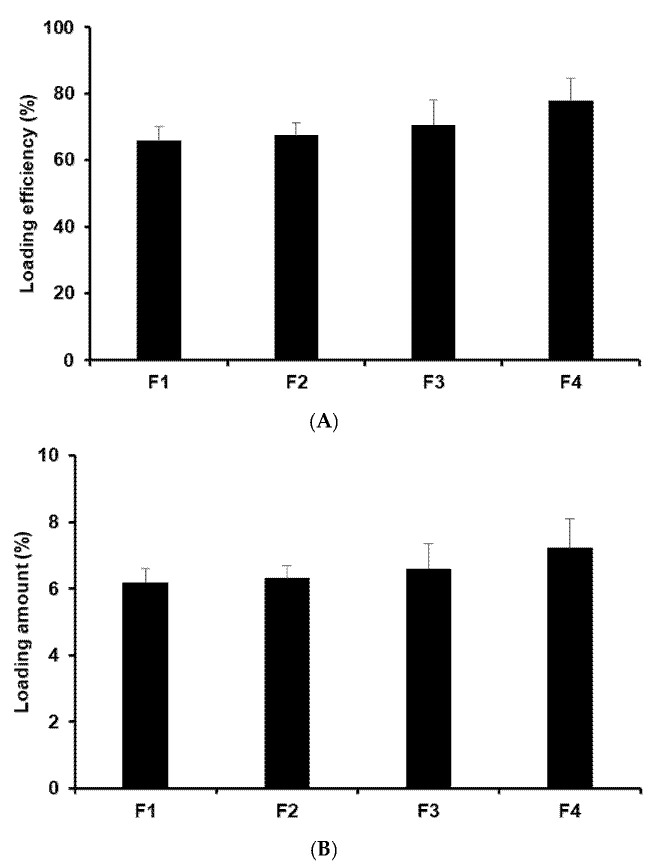
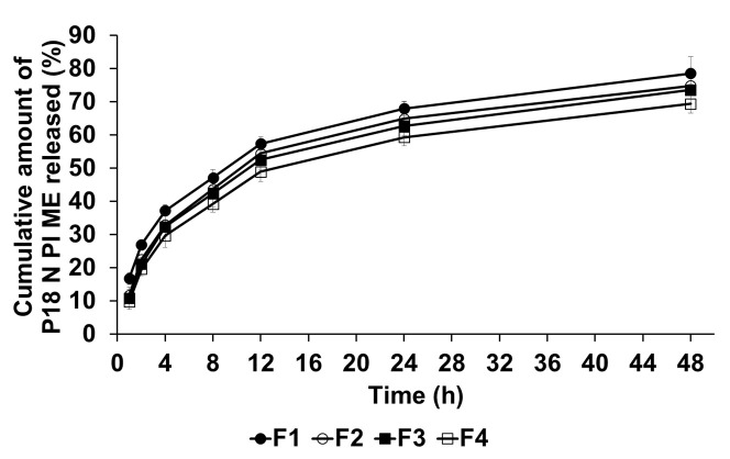
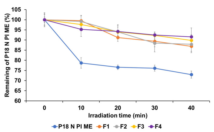
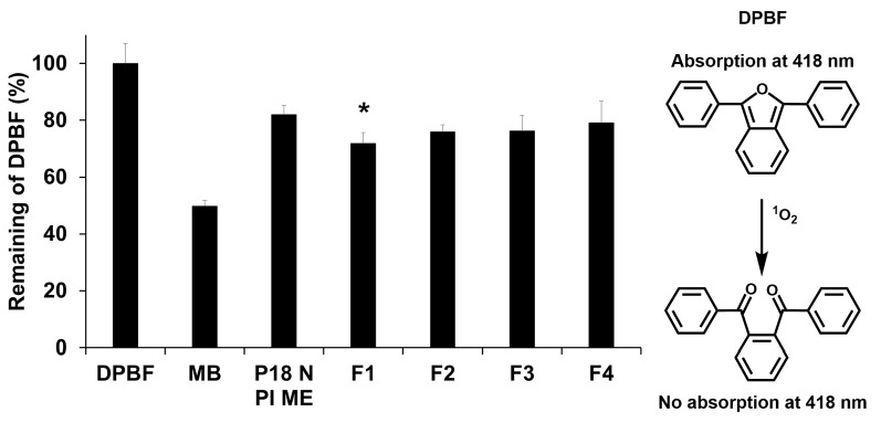
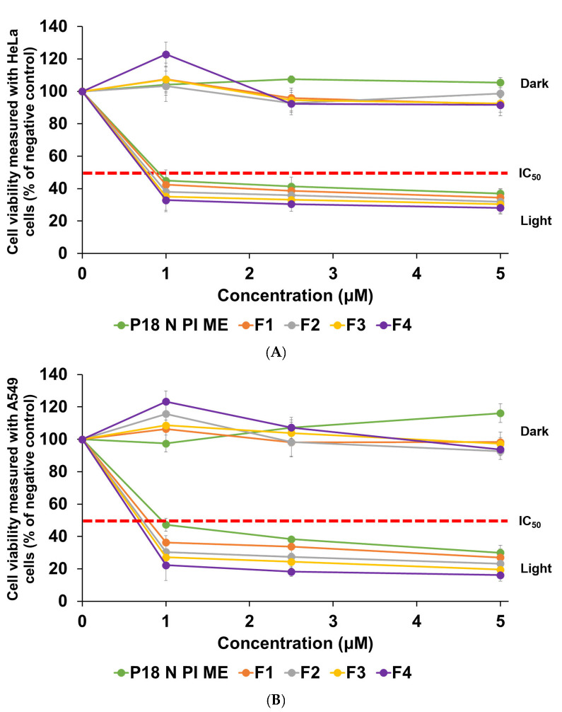
Photodynamic therapy (PDT) is an innovative cancer treatment that utilizes light. When light irradiates, purpurin-18-N-propylimide methyl ester (P18 N PI ME) generates reactive oxygen species that destroy cancer cells. The hydrophobic nature of P18 N PI ME presents challenges regarding its aggregation in the body, which can affect its effectiveness. This study aimed to enhance the bioavailability and effectiveness of cancer treatment by synthesizing P18 N PI ME and formulating P18 N PI ME-loaded solid lipid nanoparticles (SLNs). The efficacy of PDT was estimated using the 1,3-diphenylisobenzofuran (DPBF) assay and photocytotoxicity tests on the HeLa (human cervical carcinoma) and A549 (human lung carcinoma) cell lines. The P18 N PI ME-loaded SLNs demonstrated particle sizes in the range of 158.59 nm to 248.43 nm and zeta potentials in the range of -15.97 mV to -28.73 mV. These SLNs exhibited sustained release of P18 N PI ME. DPBF analysis revealed enhanced PDT effects with SLNs containing P18 N PI ME compared with standalone P18 N PI MEs. Photocytotoxicity assays indicated toxicity under light irradiation but no toxicity in the dark. Furthermore, the smallest-sized formulation exhibited the most effective photodynamic activity. These findings indicate the potential of P18 N PI ME-loaded SLNs as promising strategies for PDT in cancer therapy.

Keywords: photodynamic therapy; photosensitizers; purpurin-18-N-propylimide methyl ester; solid lipid nanoparticle.


Conflict of interest statement

The authors declare no conflicts of interest.

Figures

Figure 1
Figure 1
(A) Schematic representation of the synthesis of P18 N PI ME from P18ME; (B) 1H-NMR spectrum of P18 N PI ME (500 MHz, CDCl3, 25 °C, TMS).
Figure 2
Figure 2
UV–vis spectrum and the calibration curve of P18 N PI ME. (A) Specificity data for P18 N PI ME, placebo (no P18 N PI ME), and P18 N PI ME-loaded SLNs in MeOH at 25 °C. (B) Linearity data for the standard solution of P18 N PI ME in MeOH.
Figure 3
Figure 3
Particle characterization of P18 N PI ME-loaded SLNs manufactured using various components. (A) Particle size and polydispersity index (PDI) and (B) zeta potential. Results are presented as means ± standard deviation from three independent experiments (n = 3).
Figure 4
Figure 4
(A) Loading efficiency (LE) and (B) loading amount (LA) of P18 N PI ME in the formulations. Results are expressed as means ± standard deviation from three independent experiments (n = 3).
Figure 5
Figure 5
Cumulative release percentage profiles of P18 N PI ME from SLNs in the release medium, determined using the dialysis bag method. Results are presented as means ± standard error from three independent experiments (n = 3).
Figure 6
Figure 6
Photostability assessment of P18 N PI ME solution, using the percentage of non-degraded P18 N PI ME in both the solution and SLNs before and after LED irradiation. Results are expressed as means ± standard deviations of three independent experiments (n = 3).
Figure 7
Figure 7
Reduction rate (%) of DPBF absorbance at 418 nm for P18 N PI ME with or without SLNs after light exposure (total light dose of 2 J/cm2; exposure time of 15 min). Statistical significance of the difference in DPBF degradation between P18 N PI ME and formulation is indicated by a single asterisk (p < 0.05) or double asterisks (p < 0.01). Results are shown as means ± standard deviation for triplicates (n = 3). NC: DPBF (1,3-diphenylisobenzofuran); PC: MB (methylene blue).
Figure 8
Figure 8
Cytotoxicity of P18 N PI ME solution and formulations F1, F2, F3, and F4 against (A) HeLa and (B) A549 cell lines. The WST assay was used for the estimation of viability. Results are exhibited as means ± standard deviation for triplicates (n = 3).

Similar articles

References

    1. Warburg O. On the origin of cancer cells. Science. 1956;123:309–314. doi: 10.1126/science.123.3191.309. - DOI - PubMed
    1. Dabo-Trubelja A., Gottumukkala V. Review of cancer therapies for the perioperative physician. Perioper. Med. 2023;12:25. doi: 10.1186/s13741-023-00315-1. - DOI - PMC - PubMed
    1. Baskar R., Dai J., Wenlong N., Yeo R., Yeoh K.-W. Biological response of cancer cells to radiation treatment. Front. Mol. Biosci. 2014;1:24. doi: 10.3389/fmolb.2014.00024. - DOI - PMC - PubMed
    1. Rominiyi O., Vanderlinden A., Clenton S.J., Bridgewater C., Al-Tamimi Y., Collis S.J. Tumour treating fields therapy for glioblastoma: Current advances and future directions. Br. J. Cancer. 2021;124:697–709. doi: 10.1038/s41416-020-01136-5. - DOI - PMC - PubMed
    1. Abbas Z., Rehman S. An overview of cancer treatment modalities. Neoplasm. 2018;1:139–157.

LinkOut - more resources