. 2024 Sep 27;25(19):10391.
doi: 10.3390/ijms251910391.

Optical Nanoscopy of Cytokine-Induced Structural Alterations of the Endoplasmic Reticulum and Golgi Apparatus in Insulin-Secreting Cells

Affiliations

Optical Nanoscopy of Cytokine-Induced Structural Alterations of the Endoplasmic Reticulum and Golgi Apparatus in Insulin-Secreting Cells

Licia Anna Pugliese et al. Int J Mol Sci. .

Abstract

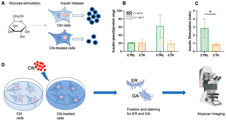
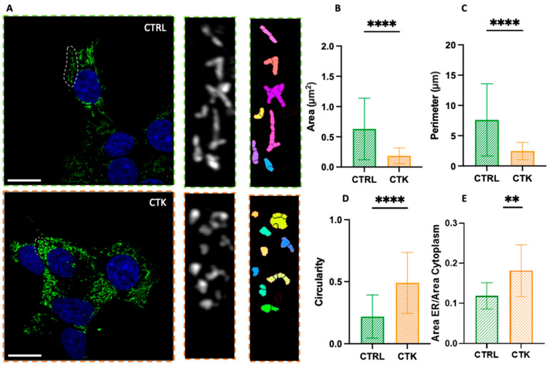
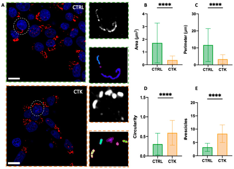
Pro-inflammatory cytokines play a role in the failure of β cells in type 1 and type 2 diabetes. While existing data from 'omics' experiments allow for some understanding of the molecular mechanisms behind cytokine-induced dysfunction in β cells, no report thus far has provided information on the direct imaging of the β cell landscape with nanoscale resolution following cytokine exposure. In this study, we use Airyscan-based optical super-resolution microscopy of Insulinoma 1E (INS-1E) cells to investigate the structural properties of two subcellular membranous compartments involved in the production, maturation and secretion of insulin-containing granules, the endoplasmic reticulum (ER) and the Golgi apparatus (GA). Our findings reveal that exposure of INS-1E cells to IL-1β and IFN-γ for 24 h leads to significant structural alterations of both compartments. In more detail, both the ER and the GA fragment and give rise to vesicle-like structures with markedly reduced characteristic area and perimeter and increased circularity with respect to the original structures. These findings complement the molecular data collected thus far on these compartments and their role in β cell dysfunction and lay the groundwork for future optical microscopy-based ex vivo and in vivo investigations.

Keywords: Airyscan; endoplasmic reticulum; fluorescence; golgi apparatus; pro-inflammatory cytokines; super resolution; β cells.


Conflict of interest statement

The authors declare no competing interests.

Figures

Figure 1
Figure 1
Schematic representation of the general workflow of our experiments. (A) After glucose stimulation, insulin secretion by INS-1E cells was evaluated. It was demonstrated that cytokine treatment impairs INS-1E insulin secretory response to glucose. (B) Effect of cytokine exposure on insulin release (corrected for protein content). (C) Effect of cytokine exposure on insulin stimulation index (ISI). Unpaired t-test, * p < 0.05. CTRL = control; CTK = cytokine; G = glucose. (D) INS-1E was plated and then incubated for 24 h in fresh complete medium or supplemented with cytokines (ctks). Then, the specimens were chemically fixed, stained with specific antibodies against the ER and GA and imaged using Airyscan microscopy (AM) (Jena, Germany).
Figure 2
Figure 2
Airyscan imaging of the ER. (A) Confocal images of the endoplasmic reticulum network, with algorithmic segmentation executed via the MorphoLibJ plugin, observed in both control and cytokine-treated INS-1E cells. Analysis of endoplasmic reticulum vesicles’ structure (see exemplary regions highlighted by the dashed circles in both CTRL and CTK images) reveals a decrease in area and perimeter ((B) and (C), respectively), along with an increased circularity value (approaching 1) (D) in samples treated with cytokines. In this analysis, regions of the cells exhibiting visible vesicles were scrutinized (number of vesicles = 128 for control and 132 for treated samples; across n = 3 independent experiments). Statistical analysis was performed using a Mann–Whitney test (**** p < 0.0001). (E) The ratio of the ER area to the cytoplasm area is greater in cells treated with cytokines. Number of cells = 12 for control and 12 for treated samples across n = 3 independent experiments. Statistical analysis was performed using a parametric t test (** p < 0.01). Scale bar indicates 10 µm.
Figure 3
Figure 3
Airyscan imaging of the GA. (A) Golgi apparatus network confocal images and algorithm segmentation in control and cytokine-treated INS-1E cells. Area, perimeter and circularity of the single vesicles are shown in the tables. The structural analysis of GA vesicles (see exemplary regions highlighted by the dashed circles in both CTRL and CTK images) shows reduced area and perimeter ((B) and (C), respectively), high circulatory value (tending to 1) (D) and higher number of vesicles (E) in cytokine-treated samples. These results support the idea of vesicle fragmentation after cytokine treatment (number of cells = 45 for control and treated samples; n = 3 independent experiments). A Mann–Whitney test was performed (**** p < 0.0001). Cells were acquired by confocal microscope using 405 and 488 excitation light, with 63X/NA1.4 objective lens. Scale bar 10 µm.

References

    1. Omar-Hmeadi M., Idevall-Hagren O. Insulin granule biogenesis and exocytosis. Cell. Mol. Life Sci. 2021;78:1957–1970. doi: 10.1007/s00018-020-03688-4. - DOI - PMC - PubMed
    1. Germanos M., Gao A., Taper M., Yau B., Kebede M.A. Inside the Insulin Secretory Granule. Metabolites. 2021;11:515. doi: 10.3390/metabo11080515. - DOI - PMC - PubMed
    1. Davidson H.W., Rhodes C.J., Hutton J.C. Intraorganellar calcium and pH control proinsulin cleavage in the pancreatic β cell via two distinct site-specific endopeptidases. Nature. 1988;333:93–96. doi: 10.1038/333093a0. - DOI - PubMed
    1. Smeekens S.P., Montag A.G., Thomas G., Albiges-Rizo C., Carroll R., Benig M., Phillips L.A., Martin S., Ohagi S., Gardner P. Proinsulin processing by the subtilisin-related proprotein convertases furin, PC2, and PC3. Proc. Natl. Acad. Sci. USA. 1992;89:8822–8826. doi: 10.1073/pnas.89.18.8822. - DOI - PMC - PubMed
    1. Orci L., Ravazzola M., Perrelet A. (Pro)insulin associates with Golgi membranes of pancreatic B cells. Proc. Natl. Acad. Sci. USA. 1984;81:6743–6746. doi: 10.1073/pnas.81.21.6743. - DOI - PMC - PubMed

MeSH terms

LinkOut - more resources