Review
. 2024 Sep 27;25(19):10401.
doi: 10.3390/ijms251910401.

Basic Guide for Approaching Drug Delivery with Extracellular Vesicles

Affiliations
Review

Basic Guide for Approaching Drug Delivery with Extracellular Vesicles

Sergey Brezgin et al. Int J Mol Sci. .

Abstract

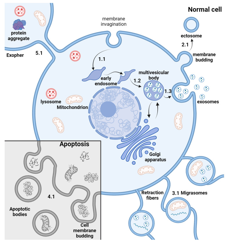
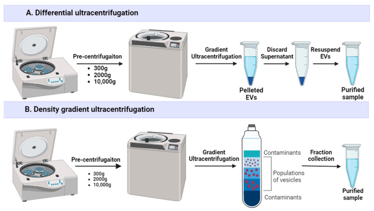
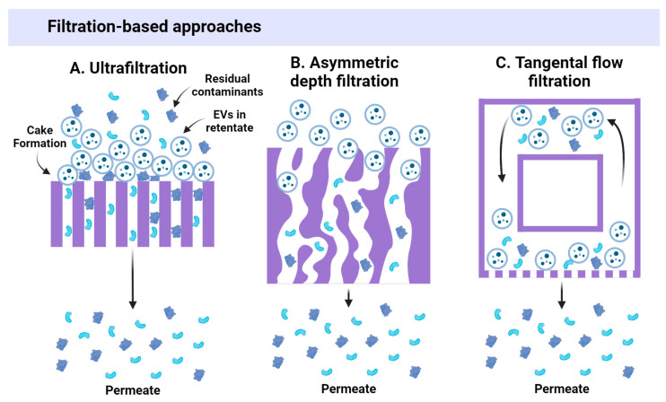
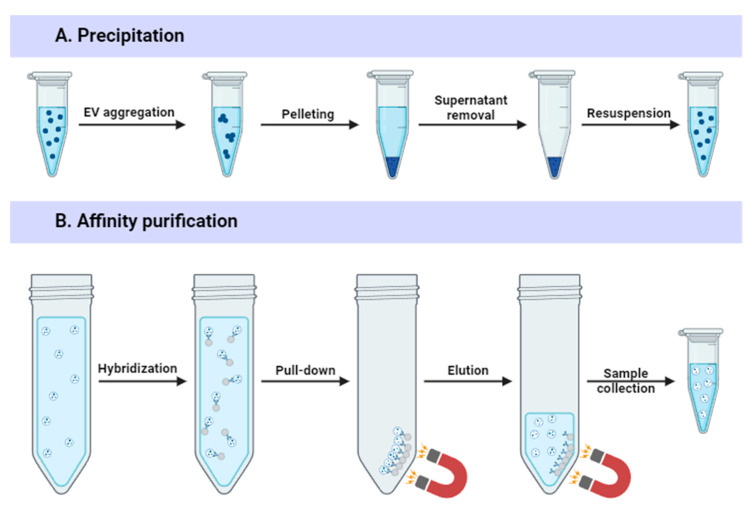
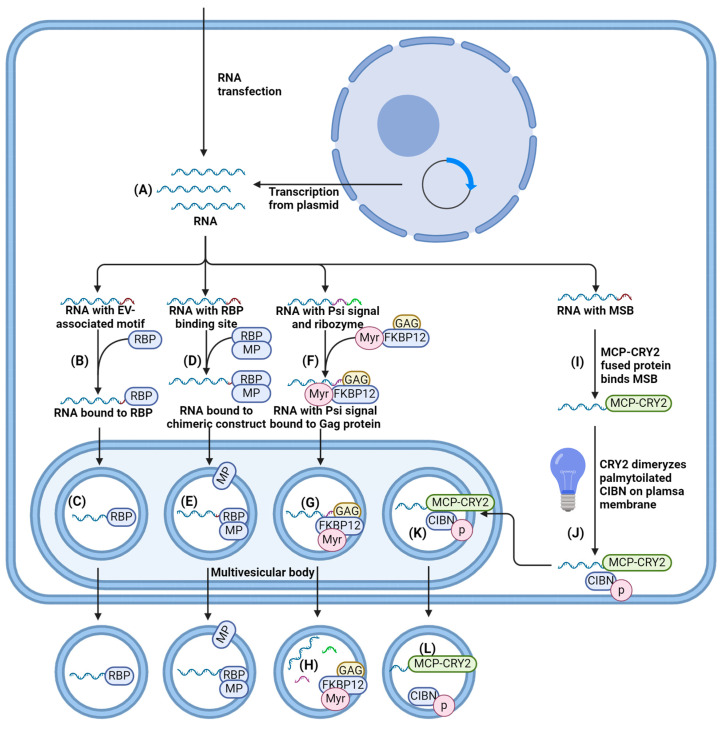
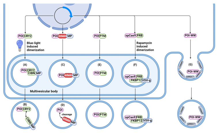
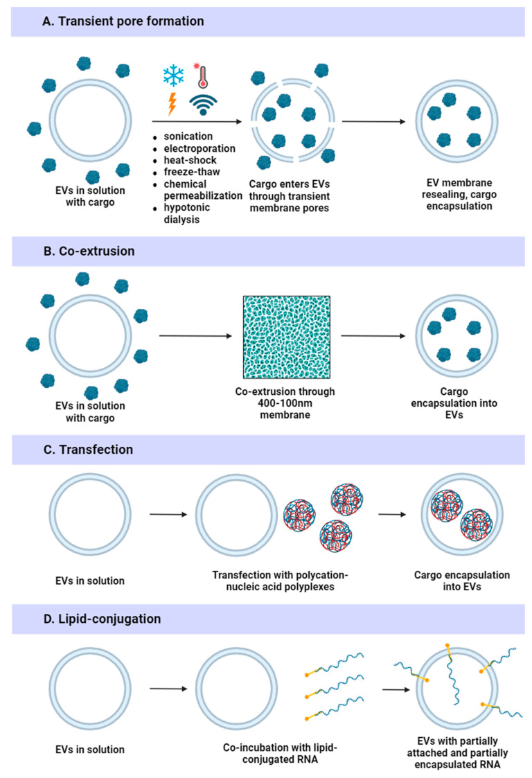
Extracellular vesicles (EVs) are natural carriers of biomolecules that play a crucial role in cell-to-cell communication and tissue homeostasis under normal and pathological conditions, including inflammatory diseases and cancer. Since the discovery of the pro-regenerative and immune-modulating properties of EVs, EV-based therapeutics have entered clinical trials for conditions such as myocardial infarction and autoimmune diseases, among others. Due to their unique advantages-such as superior bioavailability, substantial packaging capacity, and the ability to traverse biological barriers-EVs are regarded as a promising platform for targeted drug delivery. However, achieving a sufficient accumulation of therapeutic agents at the target site necessitates a larger quantity of EVs per dose compared to using EVs as standalone drugs. This challenge can be addressed by administering larger doses of EVs, increasing the drug dosage per administration, or enhancing the selective accumulation of EVs at target cells. In this review, we will discuss methods to improve the isolation and purification of EVs, approaches to enhance cargo packaging-including proteins, RNAs, and small-molecule drugs-and technologies for displaying targeting ligands on the surface of EVs to facilitate improved targeting. Ultimately, this guide can be applied to the development of novel classes of EV-based therapeutics and to overcoming existing technological challenges.

Keywords: nanotherapeutics; surface display; targeted drug delivery.


Conflict of interest statement

The authors declare no conflicts of interest.

Figures

Figure 1
Figure 1
The biogenesis of extracellular vesicles. Exosome biogenesis: 1.1 Early endosome forms from cell membrane invagination. 1.2 During the maturation process, membrane invaginations form on endosomal membrane, which subsequently bud into endosomal lumen, becoming intraluminal vesicles. 1.3 The multivesicular body fuses with the cellular membrane, releasing exosomes into the extracellular space. Ectosome biogenesis: 2.1 Ectosome biogenesis happens through direct membrane budding and fission. Migrasome biogenesis: 3.1 Migrasomes form on retraction fibers during cellular migration due to interactions with the extracellular matrix. Apoptotic bodies biogenesis: 4.1 Apoptotic bodies from the fragmented cellular membrane during apoptosis. Exopher biogenesis: Exopher forms by cell membrane budding. For a period of time it remains connected to the cell with a nanotube, but it is subsequently disconnected from the cell. 1.1 Early endosomes are formed through the invagination of the cell membrane. 1.2 During the maturation process, membrane invaginations occur on the endosomal membrane, which subsequently bud into the endosomal lumen, resulting in the formation of intraluminal vesicles. 1.3 Multivesicular bodies fuse with the cellular membrane, releasing exosomes into the extracellular space. Ectosome Biogenesis: 2.1 Ectosomes are generated through direct membrane budding and fission. Migrasome Biogenesis: 3.1 Migrasomes form on retraction fibers during cellular migration as a result of interactions with the extracellular matrix. Apoptotic Body Biogenesis: 4.1 Apoptotic bodies arise from the fragmentation of the cellular membrane during apoptosis. Exopher Biogenesis: Exophers are formed by the budding of the cell membrane. For a period, they remain connected to the cell via a nanotube but are subsequently detached from the cell.
Figure 2
Figure 2
Ultracentrifugation-based EV purification methods. (A) Differential ultracentrifugation begins with several pre-centrifugation steps to remove cells, cell debris, and large non-exosomal vesicles. Subsequently, extracellular vesicles (EVs) are pelleted using an ultracentrifuge. The supernatant, which contains contaminants, is discarded, and the EVs are resuspended in an appropriate buffer. (B) Density gradient ultracentrifugation also involves multiple pre-centrifugation steps to eliminate large contaminants. Following pre-centrifugation, the sample is applied to the density gradient, and ultracentrifugation is conducted. The fraction containing the desired EV population is then collected.
Figure 3
Figure 3
Ultrafiltration-based EV purification methods. (A) During ultrafiltration, pressure forces particles through the filter membrane. Smaller particles permeate the membrane and are discarded, while EVs cannot permeate the membrane and remain in the retentate. However, the inability of larger particles to permeate the membrane leads to the formation of a cake, which hinders the passage of residual smaller molecules through the membrane, resulting in reduced purification efficiency. (B) During asymmetric depth filtration, pressure forces the sample through a filter membrane characterized by asymmetric irregular pores. These irregular pores allow particles to enter the membrane, but their movement is impeded based on size. Smaller particles can move further or even pass through the membrane, while larger particles become entrapped and can be eluted later. (C) Tangential flow filtration is based on the continuous cyclic flow of particles through a closed system that incorporates a filter membrane into the wall of a filter cartridge. The pressure in the system drives the sample through the membrane, while the feed flow prevents cake formation.
Figure 4
Figure 4
Precipitation and affinity-based EV purification methods. (A) The addition of the cationic polymer induces EV aggregation. The sample is centrifuged, which results in EV pelleting and some of the contaminants residing in the supernatant. The supernatant is subsequently removed and EVs are resuspended in a suitable buffer. (B) During affinity purification, antibodies against EV markers conjugated to the magnetic beads are added to the sample. After the binding of antibodies, the beads are pulled down by magnetic force. The supernatant is replaced by the elution buffer, resulting in the release of the EVs. Afterward, the sample is collected.
Figure 5
Figure 5
Endogenous RNA loading methods. (A) RNA is delivered into the cell via transfection with RNA or a plasmid encoding the RNA. (B) The EV-associated motif in RNA facilitates the binding of EV-associated RNA-binding proteins (RBPs). (C) EV-associated RBPs assist in the sorting of RNA into extracellular vesicles (EVs). (D) A chimeric construct consisting of an RBP and an EV membrane protein (MP) binds to the RBP-binding site in RNA. (E) The RBP-MP complex bound to RNA is sorted to the EV membrane. (F) A chimeric protein consisting of myristoylated FKBP12 and GAG binds to the Psi signal in RNA. (G) Due to myristoylation, the RNA-bound construct is sorted into EVs. (H) A ribozyme encoded in the RNA induces self-cleavage and the release of the RNA. (I) MCP fused to CRY2 binds to the MSB site in the RNA. (J) Blue light induces the dimerization of CRY2 with palmitoylated CIBN, which is enriched on the cell membrane. (K) The dimer bound to RNA is sorted into the EV. (L) In the absence of blue light, the dimer disassembles.
Figure 6
Figure 6
Endogenous protein loading methods. (A) The protein of interest (POI) is fused with CRY2. Upon exposure to blue light, the POI-CRY2 complex dimerizes with CIBN, which is fused to an EV membrane protein (MP). (B) In the absence of blue light, the dimer disassembles, releasing the protein into the EV lumen. (C) The chimeric protein, consisting of the POI, a self-cleaving intein, and the MP, is sorted into the EV. (D) The intein cleaves the construct, releasing the POI into the EV lumen. (E) Post-translational modifications (PTMs), such as myristoylation or palmitoylation, facilitate the sorting of the protein into EVs. (F) Rapamycin induces the dimerization of SpCas9-FRB with FKBP-VSV-g. Due to VSV-g, the construct is sorted into the EV. (G) The POI, fused to a WW signal, binds to ARRDC1, leading to their sorting into ARRDC1-mediated microvesicles.
Figure 7
Figure 7
Exogenous cargo loading methods. (A) Several methods, including sonication, electroporation, freeze-thaw cycles, heat shock, chemical permeabilization, and hypotonic dialysis, induce transient pore formation in membranes. Through these pores, cargo from the extravesicular space can enter the vesicle. Subsequently, the membrane reseals, trapping the cargo inside. (B) Co-extrusion leads to a transient decrease in membrane continuity, facilitating the entry of cargo into EVs. (C) Similar to cells, EVs can be transfected with nucleic acids using cationic transfection reagents. (D) Cargo can be conjugated to lipids and subsequently incorporated into the EV membrane; however, the cargo may exist both within the vesicle and on its surface.

Similar articles

Cited by

References

    1. Ginini L., Billan S., Fridman E., Gil Z. Insight into extracellular vesicle-cell communication: From cell recognition to intracellular fate. Cells. 2022;11:1375. doi: 10.3390/cells11091375. - DOI - PMC - PubMed
    1. Welsh J.A., Goberdhan D.C.I., O’Driscoll L., Buzas E.I., Blenkiron C., Bussolati B., Cai H., Di Vizio D., Driedonks T.A.P., Erdbrügger U. Minimal information for studies of extracellular vesicles (MISEV2023): From basic to advanced approaches. J. Extracell. Vesicles. 2024;13:e12404. doi: 10.1002/jev2.12404. - DOI - PMC - PubMed
    1. Cecchin R., Troyer Z., Witwer K., Morris K. V Extracellular vesicles: The next generation in gene therapy delivery. Mol. Ther. 2023;31:1225–1230. doi: 10.1016/j.ymthe.2023.01.021. - DOI - PMC - PubMed
    1. Banks W.A., Sharma P., Bullock K.M., Hansen K.M., Ludwig N., Whiteside T.L. Transport of extracellular vesicles across the blood-brain barrier: Brain pharmacokinetics and effects of inflammation. Int. J. Mol. Sci. 2020;21:4407. doi: 10.3390/ijms21124407. - DOI - PMC - PubMed
    1. El Andaloussi S., Mäger I., Breakefield X.O., Wood M.J.A. Extracellular vesicles: Biology and emerging therapeutic opportunities. Nat. Rev. Drug Discov. 2013;12:347–357. doi: 10.1038/nrd3978. - DOI - PubMed

LinkOut - more resources