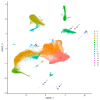
Cerebrospinal Fluid and Peripheral Blood Lymphomonocyte Single-Cell Transcriptomics in a Subject with Multiple Sclerosis Acutely Infected with HIV
- PMID: 39408789
- PMCID: PMC11476486
- DOI: 10.3390/ijms251910459
Cerebrospinal Fluid and Peripheral Blood Lymphomonocyte Single-Cell Transcriptomics in a Subject with Multiple Sclerosis Acutely Infected with HIV
Abstract
Signatures of neurodegeneration in clinical samples from a subject with multiple sclerosis (MS) acutely infected with HIV were investigated with single-cell transcriptomics using 10X Chromium technology. Sequencing was carried out on NovaSeq-TM, and the analysis was performed with Cell Ranger software (v 7.1.0) associated with a specifically established bioinformatic pipeline. A total of 1446 single-cell transcriptomes in cerebrospinal fluid (CSF) and 4647 in peripheral blood mononuclear cells (PBMCs) were obtained. In the CSF, many T-cell lymphocytes with an enriched amount of plasma cells and plasmacytoid dendritic (pDC) cells, as compared to the PBMCs, were detected. An unsupervised cluster analysis, putting together our patient transcriptomes with those of a publicly available MS scRNA-seq dataset, showed up-regulated microglial neurodegenerative gene expression in four clusters, two of which included our subject's transcriptomes. A few HIV-1 transcripts were found only in the CD4 central memory T-cells of the CSF compartment, mapping to the gag-pol, vpu, and env regions. Our data, which describe the signs of neurodegenerative gene expression in a very peculiar clinical situation, did not distinguish the cause between multiple sclerosis and HIV infection, but they can give a glimpse of the high degree of resolution that may be obtained by the single-cell transcriptomic approach.
Keywords: immune dysregulation in infectious diseases; interactions between infective agents and immune responses.
Conflict of interest statement
The authors declare that they have no known competing financial interests or personal relationships that could have appeared to influence the work reported in this paper.
Figures


References
-
- Valcour V., Chalermchai T., Sailasuta N., Marovich M., Lerdlum S., Suttichom D., Suwanwela N.C., Jagodzinski L., Michael N., Spudich S., et al. Central Nervous System Viral Invasion and Inflammation During Acute HIV Infection. J. Infect. Dis. 2012;206:275–282. doi: 10.1093/infdis/jis326. - DOI - PMC - PubMed
-
- Souza F.d.S., Freitas N.L., Gomes Y.C.P., Torres R.C., Echevarria-Lima J., da Silva-Filho I.L., Leite A.C.C.B., de Lima M.A.S.D., da Silva M.T.T., Araújo A.d.Q.C., et al. Following the Clues: Usefulness of Biomarkers of Neuroinflammation and Neurodegeneration in the Investigation of HTLV-1-Associated Myelopathy Progression. Front. Immunol. 2021;12:737941. doi: 10.3389/fimmu.2021.737941. - DOI - PMC - PubMed
MeSH terms
Grants and funding
LinkOut - more resources
Full Text Sources
Medical
Molecular Biology Databases
Research Materials

