Review
. 2024 Sep 29;25(19):10503.
doi: 10.3390/ijms251910503.

The Use of Patient-Derived Organoids in the Study of Molecular Metabolic Adaptation in Breast Cancer

Affiliations
Review

The Use of Patient-Derived Organoids in the Study of Molecular Metabolic Adaptation in Breast Cancer

Natalija Glibetic et al. Int J Mol Sci. .

Abstract

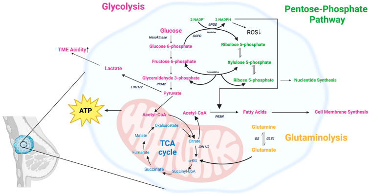
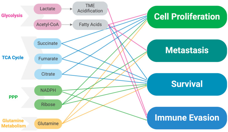
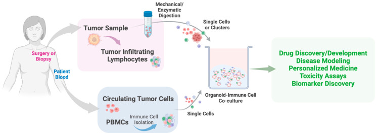
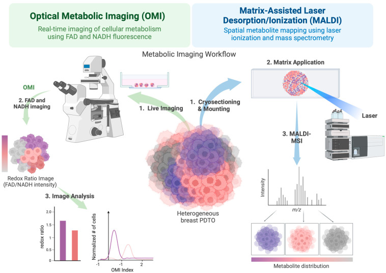
Around 13% of women will likely develop breast cancer during their lifetime. Advances in cancer metabolism research have identified a range of metabolic reprogramming events, such as altered glucose and amino acid uptake, increased reliance on glycolysis, and interactions with the tumor microenvironment (TME), all of which present new opportunities for targeted therapies. However, studying these metabolic networks is challenging in traditional 2D cell cultures, which often fail to replicate the three-dimensional architecture and dynamic interactions of real tumors. To address this, organoid models have emerged as powerful tools. Tumor organoids are 3D cultures, often derived from patient tissue, that more accurately mimic the structural and functional properties of actual tumor tissues in vivo, offering a more realistic model for investigating cancer metabolism. This review explores the unique metabolic adaptations of breast cancer and discusses how organoid models can provide deeper insights into these processes. We evaluate the most advanced tools for studying cancer metabolism in three-dimensional culture models, including optical metabolic imaging (OMI), matrix-assisted laser desorption/ionization mass spectrometry imaging (MALDI-MSI), and recent advances in conventional techniques applied to 3D cultures. Finally, we explore the progress made in identifying and targeting potential therapeutic targets in breast cancer metabolism.

Keywords: MALDI-MSI; breast cancer; glycolysis; lipid metabolism; metabolism; optical metabolic imaging; organoid; tumor microenvironment.


Conflict of interest statement

The authors declare no conflicts of interest. The funders had no role in the design of the study; in the collection, analyses, or interpretation of data; in the writing of the manuscript; or in the decision to publish the results.

Figures

Figure 1
Figure 1
A non-exhaustive representation of key metabolic pathway alterations in breast cancer cells, including glycolysis, the TCA cycle, the pentose phosphate pathway (PPP), and glutaminolysis. Breast cancer cells show increased glycolysis, converting glucose to lactate (Warburg effect), contributing to an acidic TME. Key enzymes such as hexokinase (HK), pyruvate kinase M2 (PKM2), and lactate dehydrogenase (LDH1/2) are involved. The oxidative PPP is upregulated, increasing ribose-5-phosphate and NADPH production. Alterations in TCA cycle enzymes (IDH1/2) and glutamine metabolism (GLS1) support biosynthesis and proliferation. Fatty acid synthesis is upregulated, with fatty acid synthase (FASN) producing fatty acids from acetyl-CoA. Created with BioRender.com.
Figure 2
Figure 2
Metabolite influence on cancer cell behavior. Key metabolites from glycolysis, TCA cycle, PPP, and glutamine metabolism and their roles in driving cancer cell processes. Metabolites from these pathways contribute to cell proliferation, metastasis, survival, and immune evasion. Lactate and acetyl-CoA (glycolysis) promote TME acidification and fatty acid synthesis. Succinate, fumarate, and citrate (TCA cycle) support growth and survival. NADPH and ribose (PPP) are involved in biosynthesis and immune evasion, while glutamine fuels multiple processes. Colored arrows indicate specific metabolite influences on cellular behaviors. Created with BioRender.com.
Figure 3
Figure 3
Schematic representation of PDTO-immune cell co-culture workflow and functional testing. PDTO-immune cell co-culturing begins with sourcing and digesting patient tumor samples to isolate clusters or single cancer cells and tumor-infiltrating lymphocytes (TILs) or by isolating circulating tumor cells and PBMCs from patient blood. The cells are then embedded in an ECM for structural support. Fully formed tumor organoids co-cultured with immune cells (macrophages, T cells, B cells, NK cells, etc.) may be used in drug discovery, personalized medicine, therapeutic toxicity studies, and biomarker discovery. Created with BioRender.com.
Figure 4
Figure 4
Metabolic imaging of breast cancer PDTOs using OMI and MALDI-MSI. Schematic representation of imaging workflow for assessing metabolic activity in breast cancer PDTOs using OMI and MALDI-MSI. In the OMI workflow (left), autofluorescence of NADH and FAD is used to monitor cellular metabolism. This involves the acquisition of live images (1) and generating a redox ratio image (2) that reflects the metabolic state of the cells. Finally, image analysis is performed to compute the redox ratio and OMI index, quantifying metabolic heterogeneity across the PDTO. In the MALDI-MSI workflow (right), PDTOs are prepared for mass spectrometry analysis. First, organoids are cryosectioned and mounted (1), followed by matrix application (2) to facilitate laser-based ionization. MALDI-mass spectrometry imaging is conducted (3), producing an m/z spectrum and spatially resolved metabolic heatmaps, illustrating metabolite distribution within the PDO. The integration of OMI and MALDI-MSI enables high-resolution metabolic profiling of PDOs, providing insights into tumor metabolic heterogeneity and potential therapeutic targets. Created with BioRender.com.

Similar articles

Cited by

References

    1. National Cancer Institute Surveillance, Epidemiology, and End Results Program. Breast Cancer—Breast Cancer Risk from Birth over Time, by Sex, All Races/Ethnicities, Risk of Being Diagnosed with Cancer (2018–2019, 2021) [(accessed on 23 August 2024)]; Available online: https://seer.cancer.gov/statistics-network/explorer/
    1. Centers for Disease Control and Prevention. U.S. Department of Health and Human Services U.S. Cancer Statistics Female Breast Cancer Stat Bite. [(accessed on 23 August 2024)];2024 Available online: https://www.cdc.gov/united-states-cancer-statistics/publications/breast-....
    1. Pavlova N.N., Thompson C.B. The Emerging Hallmarks of Cancer Metabolism. Cell Metab. 2017;23:27. doi: 10.1016/j.cmet.2015.12.006. - DOI - PMC - PubMed
    1. Warburg O. On the Origin of Cancer Cells. Science. 1956;123:309–314. doi: 10.1126/science.123.3191.309. - DOI - PubMed
    1. Warburg O., Wind F., Negelein E. The Metabolism of Tumors in the Body. J. Gen. Physiol. 1927;8:519–530. doi: 10.1085/jgp.8.6.519. - DOI - PMC - PubMed

LinkOut - more resources