. 2024 Sep 29;25(19):10506.
doi: 10.3390/ijms251910506.

Bovine Neutrophil β-Defensin-5 Provides Protection against Multidrug-Resistant Klebsiella pneumoniae via Regulating Pulmonary Inflammatory Response and Metabolic Response

Affiliations

Bovine Neutrophil β-Defensin-5 Provides Protection against Multidrug-Resistant Klebsiella pneumoniae via Regulating Pulmonary Inflammatory Response and Metabolic Response

Shuxin Zhu et al. Int J Mol Sci. .

Abstract

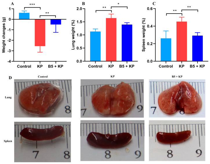
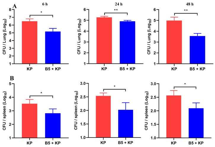
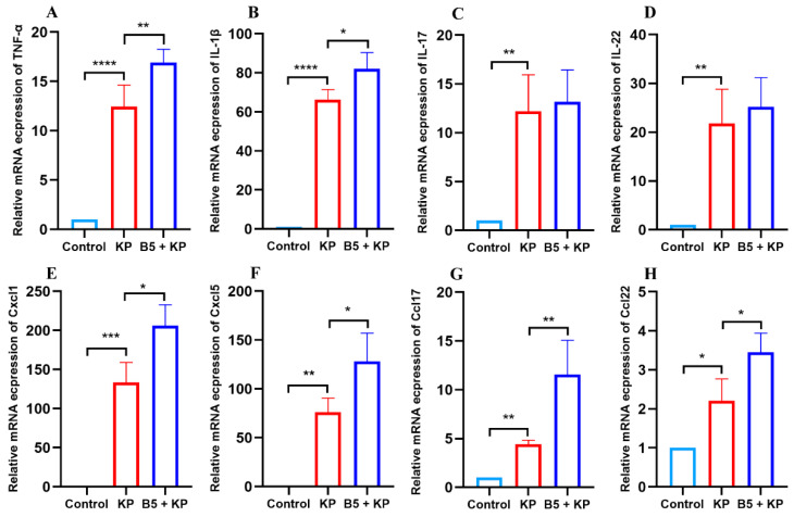
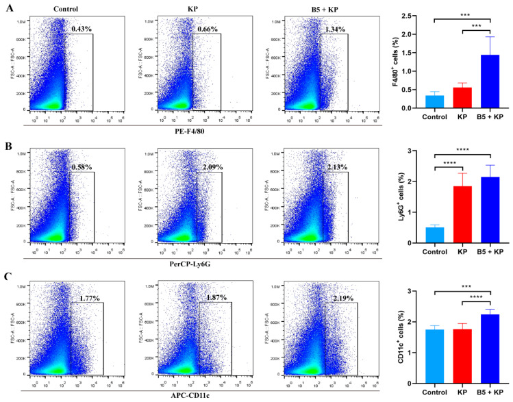
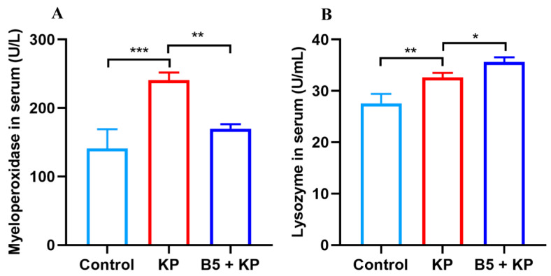
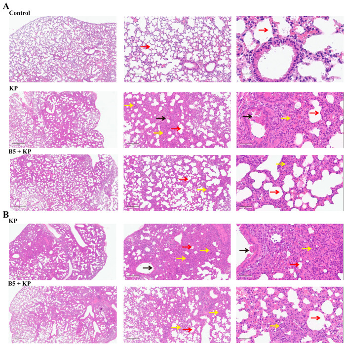
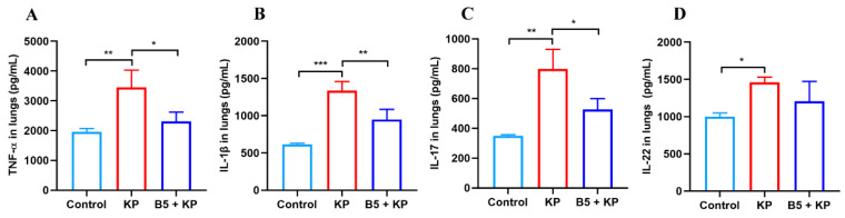
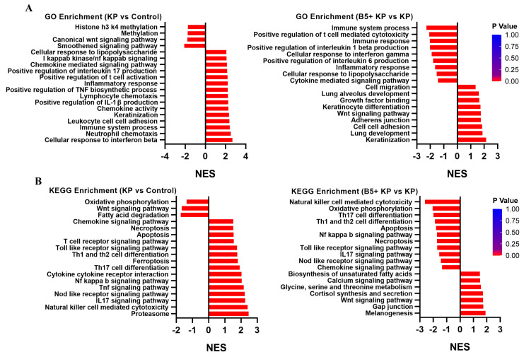
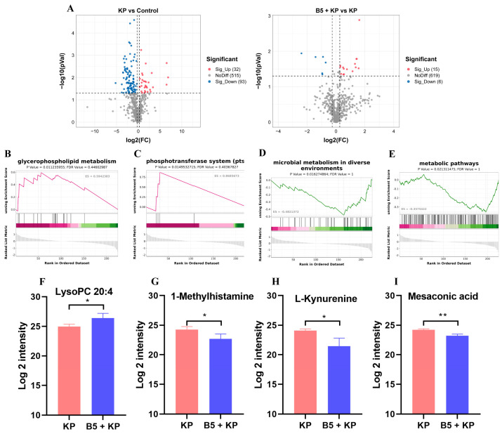
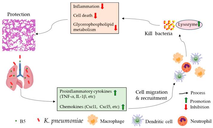
Klebsiella pneumoniae (K. pneumoniae), a kind of zoonotic bacteria, is among the most common antibiotic-resistant pathogens, and it causes nosocomial infections that pose a threat to public health. In this study, the roles of synthetic bovine neutrophil β-defensin-5 (B5) in regulating inflammatory response and metabolic response against multidrug-resistant K. pneumoniae infection in a mouse model were investigated. Mice were administrated intranasally with 20 μg of B5 twice and challenged with K. pneumoniae three days after B5 pretreatment. Results showed that B5 failed to directly kill K. pneumoniae in vitro, but it provided effective protection against multidrug-resistant K. pneumoniae via decreasing the bacterial load in the lungs and spleen, and by alleviating K. pneumoniae-induced histopathological damage in the lungs. Furthermore, B5 significantly enhanced the mRNA expression of TNF-α, IL-1β, Cxcl1, Cxcl5, Ccl17, and Ccl22 and obviously enhanced the rapid recruitment of macrophages and dendritic cells in the lungs in the early infection phase, but significantly down-regulated the levels of TNF-α, IL-1β, and IL-17 in the lungs in the later infection phase. Moreover, RNA-seq results showed that K. pneumoniae infection activated signaling pathways related to natural killer cell-mediated cytotoxicity, IL-17 signaling pathway, inflammatory response, apoptosis, and necroptosis in the lungs, while B5 inhibited these signaling pathways. Additionally, K. pneumoniae challenge led to the suppression of glycerophospholipid metabolism, the phosphotransferase system, the activation of microbial metabolism in diverse environments, and metabolic pathways in the lungs. However, B5 significantly reversed these metabolic responses. Collectively, B5 can effectively regulate the inflammatory response caused by K. pneumoniae and offer protection against K. pneumoniae. B5 may be applied as an adjuvant to the existing antimicrobial therapy to control multidrug-resistant K. pneumoniae infection. Our study highlights the potential of B5 in enhancing pulmonary bacterial clearance and alleviating K. pneumoniae-caused inflammatory damage.

Keywords: Klebsiella pneumoniae; bovine neutrophil β-defensin-5; inflammatory response; metabolic response.


Conflict of interest statement

The authors declare no conflicts of interest.

Figures

Figure 1
Figure 1
B5 ameliorates K. pneumoniae-caused weight loss and organ enlargement. Mice were treated intranasally with B5 (20 μg) and challenged with K. pneumoniae three days after treatment and euthanized at 24 h after challenge. (A) Body weight changes. (B) Weight of lung tissues. (C) Weight of spleen tissues. (D) Gross pathology of lungs and spleen. Data shown are means ± SD. Data are representative of two independent experiments (n = 8 mice per group). Statistical analysis was performed by ANOVA followed by Tukey’s multiple comparison test (* p < 0.033, ** p < 0.0021, *** p < 0.0002).
Figure 2
Figure 2
B5 reduces bacterial load in lungs and spleen. Lung and spleen tissues were collected at 6 h, 24 h, and 48 h after challenge. (A) Bacterial load in lungs. (B) Bacterial load in spleen. Data shown are means ± SD. Data are representative of two independent experiments (n = 8 mice per group). Statistical analysis was performed by ANOVA followed by Tukey’s multiple comparison test (* p < 0.033, ** p < 0.0021).
Figure 3
Figure 3
B5 up-regulates pulmonary mRNA expressions of cytokines and chemokines in early infection phase. Lung and spleen tissues were collected at 4 h after challenge. (AD) The mRNA expression levels of TNF-α (A), IL-1β (B), IL-17 (C), and IL-22 (D) in lungs. (EH) The mRNA expression levels of Cxcl1 (E), Cxcl5 (F), Ccl17 (G), and Ccl22 (H) in lungs. Data shown are means ± SD. Data are representative of two independent experiments (n = 3). Statistical analysis was performed by ANOVA followed by Tukey’s multiple comparison test (* p < 0.033, ** p < 0.0021, *** p < 0.0002, **** p < 0.0001).
Figure 4
Figure 4
B5 promotes the recruitment of innate immune cells to the lungs in the early infection phase. The lungs were collected at 6 h after challenge. (A) Percentage of macrophages (F4/80+ cells) in the lungs. (B) Percentage of neutrophils (Ly6G+ cells) in the lungs. (C) Percentage of dendritic cells (CD11c+ cells) in the lungs. Data shown are means ± SD. Data are representative of two independent experiments (n = 8 mice per group). Statistical analysis was performed by ANOVA followed by Tukey’s multiple comparison test (*** p < 0.0002, **** p < 0.0001).
Figure 5
Figure 5
B5 regulates levels of myeloperoxidase and lysozyme in serum in later infection phase. Serum samples were collected at 24 h after challenge. (A) The level of myeloperoxidase in serum. (B) The level of lysozyme in serum. Data shown are means ± SD. Data are representative of two independent experiments (n = 3). Statistical analysis was performed by ANOVA followed by Tukey’s multiple comparison test (* p < 0.033, ** p < 0.0021, *** p < 0.0002).
Figure 6
Figure 6
B5 alleviates inflammatory damage in lungs. (A) Histopathological images of lungs at 24 h after challenge. (B) Histopathological images of lungs at 48 h after challenge. Left images at ×40 magnification (scale bar, 500 μm); middle images at ×100 magnification (scale bar, 200 μm); right images at ×400 magnification (scale bar, 50 μm). Red arrows represent alveoli, black arrows represent fallen bronchial epithelial cells, and yellow arrows represent inflammatory cells in pulmonary interstitium. Data are representative of two independent experiments (n = 3 mice per group).
Figure 7
Figure 7
B5 decreases pro-inflammatory cytokine secretion in the lungs in the later stage of K. pneumoniae infection. (AD) Levels of TNF-α, IL-1β, IL-22, and IL-17 in the lungs at 24 h after challenge. Data shown are means ± SD. Data are representative of two independent experiments (n = 3). Statistical analysis was performed by ANOVA followed by Tukey’s multiple comparison test (* p < 0.033, ** p < 0.0021, *** p < 0.0002).
Figure 8
Figure 8
Gene Ontology (GO) term and Kyoto Encyclopedia of Genes and Genomes (KEGG) pathway analysis of differentially expressed genes. (A) Differentially expressed genes are shown in volcano plots. (B) Fold changes in representative differentially expressed genes. (CE) Gene enrichment in biological process (C), cellular component (D), and molecular function (E). (F) GO enrichment scatterplot. (G) KEGG enrichment scatterplot.
Figure 9
Figure 9
Gene set enrichment analysis (GSEA) of differentially expressed genes. (A) GSEA of Gene Ontology (GO) enrichment. (B) GSEA of Kyoto Encyclopedia of Genes and Genomes (KEGG) enrichment.
Figure 10
Figure 10
B5 regulates K. pneumoniae-induced metabolic response in lungs. (A) Differentially expressed metabolites are shown in volcano plots. (BE) Gene set enrichment analysis (GSEA) of KEGG enrichment (B5 + KP group vs. KP group). (FI) Representative differentially expressed metabolites. Statistical analysis was performed by Student’s t-test (* p < 0.05, ** p < 0.005).
Figure 11
Figure 11
The mechanism and mode of action of B5 against K. pneumoniae infection.

Similar articles

References

    1. Antimicrobial Resistance Collaborators Global burden of bacterial antimicrobial resistance in 2019: A systematic analysis. Lancet. 2022;399:629–655. doi: 10.1016/S0140-6736(21)02724-0. - DOI - PMC - PubMed
    1. Ye C., Li W., Yang Y., Liu Q., Li S., Zheng P., Zheng X., Zhang Y., He J., Chen Y., et al. Inappropriate use of antibiotics exacerbates inflammation through OMV-induced pyroptosis in MDR Klebsiella pneumoniae infection. Cell Rep. 2021;36:109750. doi: 10.1016/j.celrep.2021.109750. - DOI - PubMed
    1. Santacroce L., Di Domenico M., Montagnani M., Jirillo E. Antibiotic Resistance and Microbiota Response. Curr. Pharm. Des. 2023;29:356–364. doi: 10.2174/1381612829666221219093450. - DOI - PubMed
    1. O’neill J. Review on Antimicrobial Resistance. Tackling a Global Health Crisis: Rapid Diagnostics: Stopping Unnecessary Use of Antibiotics. Indep. Rev. AMR. 2015:1–36.
    1. Karami-Zarandi M., Rahdar H.A., Esmaeili H., Ranjbar R. Klebsiella pneumoniae: An update on antibiotic resistance mechanisms. Future Microbiol. 2023;18:65–81. doi: 10.2217/fmb-2022-0097. - DOI - PubMed

LinkOut - more resources