Review
. 2024 Oct 1;25(19):10584.
doi: 10.3390/ijms251910584.

Recombinant Influenza A Viruses Expressing Reporter Genes from the Viral NS Segment

Affiliations
Review

Recombinant Influenza A Viruses Expressing Reporter Genes from the Viral NS Segment

Luis Martinez-Sobrido et al. Int J Mol Sci. .

Abstract

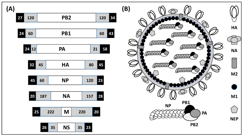
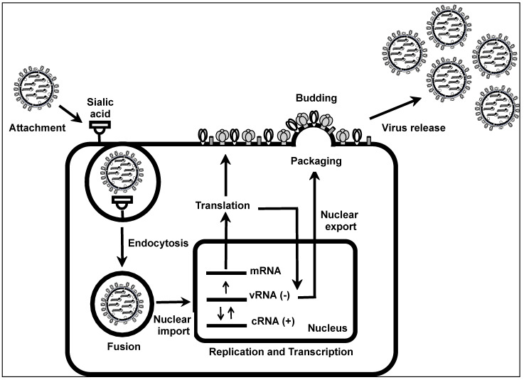
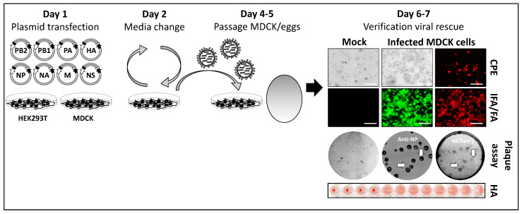
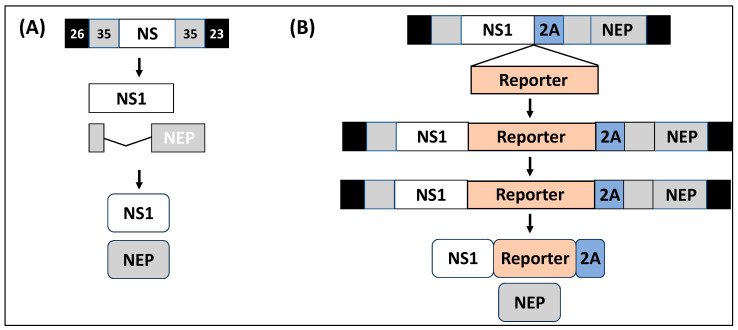
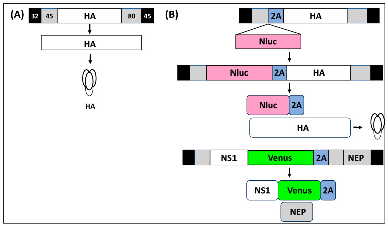
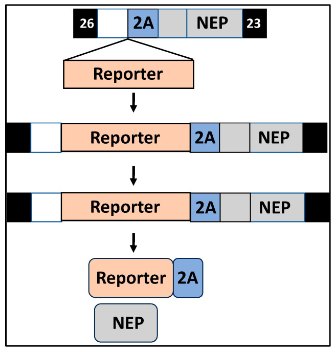
Studying influenza A viruses (IAVs) requires secondary experimental procedures to detect the presence of the virus in infected cells or animals. The ability to generate recombinant (r)IAV using reverse genetics techniques has allowed investigators to generate viruses expressing foreign genes, including fluorescent and luciferase proteins. These rIAVs expressing reporter genes have allowed for easily tracking viral infections in cultured cells and animal models of infection without the need for secondary approaches, representing an excellent option to study different aspects in the biology of IAV where expression of reporter genes can be used as a readout of viral replication and spread. Likewise, these reporter-expressing rIAVs provide an excellent opportunity for the rapid identification and characterization of prophylactic and/or therapeutic approaches. To date, rIAV expressing reporter genes from different viral segments have been described in the literature. Among those, rIAV expressing reporter genes from the viral NS segment have been shown to represent an excellent option to track IAV infection in vitro and in vivo, eliminating the need for secondary approaches to identify the presence of the virus. Here, we summarize the status on rIAV expressing traceable reporter genes from the viral NS segment and their applications for in vitro and in vivo influenza research.

Keywords: NS segment; fluorescence; luminescence; plasmid-based reverse genetics; recombinant influenza A virus; replication-competent reporter-expressing influenza A virus; reporter genes.


Conflict of interest statement

The authors declare no conflicts of interest.

Figures

Figure 1
Figure 1
IAV genome organization and viral particle structure. (A) Genomic organization: The IAV genome is made of eight negative-sense, single-stranded vRNA segments. Each viral segment contains the 3′ and 5′ non-coding regions (NCRs) involved in viral genome replication and gene transcription (black boxes). Together with the NCR, the 3′ and 5′ ends of the vRNA coding regions (gray boxes) contain the packaging signals required for efficient incorporation of the vRNAs into the new virions. The genome of IAV is organized based on the length of the vRNAs as PB2, PB1, PA, HA, NP, NA, M, and NS. (B) Viral particle structure: The surface of the influenza viral particle is made of a lipid bilayer decorated with the three major surface proteins: HA, NA, and M2. Under the viral membrane is the M1 protein. Inside the viral particle are located the NEP and the vRNAs encapsidated by the viral NP. Each of the vRNAs contains the three components of the viral RdRp subunits (PB2, PB1, and PA).
Figure 2
Figure 2
Life cycle of IAV: The different steps of the IAV life cycle include the attachment of the virus into susceptible cells, a process mediated by the interaction of IAV HA to sialic acid-containing receptors. Next, IAV is internalized into the cytoplasm of the cell using an endocytosis-mediated mechanism. Fusion of the viral membrane with the membrane of the endosome, a process mediated by the viral HA and M2, results in the release of vRNPs into the cytoplasm of infected cells. Nuclear import of vRNPs is mediated by the interaction of NLS present in the viral polymerase proteins and NP with importin α and β. Inside the nucleus, viral transcription and replication mediated by PB2, PB1, PA, and NP results in the formation of mRNAs that are translated in the cytoplasm of infected cells to produce viral proteins and the formation of complementary (c)RNAs that are used as a template to produce new vRNAs, respectively. Nuclear export of newly synthesized vRNAs is mediated by the interaction of NEP with Crm1 and M1. Inside the cytoplasm, vRNPs are selectively incorporated into nascent virions using specific packaging signals present in the 3′ and 5′ NCRs of each of the vRNAs. Budding of IAV is mediated by the interaction of M1 with newly synthesized vRNPs and viral glycoproteins in the membrane of infected cells. Finally, release of IAV from infected cells is mediated by cleavage of HA from sialic acid-containing glycoproteins in the cell membrane by NA.
Figure 3
Figure 3
Plasmid-based reverse genetics to generate rIAV: Conventional reverse genetics to generate rIAV is based on the use of 8 plasmid DNAs. Each of the viral plasmids uses an ambisense strategy to encode the vRNAs and the viral proteins under Pol I and Pol II promoters, respectively. To rescue rIAVs, the 8 ambisense plasmids are co-transfected into co-cultures of 293T and MDCK cells (Day 1). After transfection, the cell culture supernatant is replaced by fresh media containing TPCK-trypsin (Day 2). At 2–3 days after transfection, the cell culture supernatant is collected and used to infect new MDCK cells or 8–10 chicken embryonated eggs (Day 4–5). Then, 2–3 days after infection, cell culture supernatant from infected MDCK cells or the allantoic fluid from chicken embryonated eggs is harvested to confirm the presence of rIAV. Detection of rIAV can be determined by evaluating cytopathic effect (CPE); immunofluorescence (IFA), using IAV-specific antibodies, or fluorescence assay (FA) if fluorescent-expressing viruses are rescued. Plaque assay with/without immunostaining with IAV-specific antibodies and fluorescence analysis is used to confirm expression of the reporter gene from all the recombinant viruses in the preparation (arrows indicate correlation between IAV viral NP and mCherry expression). Alternatively, a conventional hemagglutination assay (HA) can be used to demonstrate the presence of rIAV in the cell culture supernatants or allantoic fluid from MDCK and eggs, respectively [8,61]. IFA/FA scale bars 200 µm.
Figure 4
Figure 4
Modified IAV NS segment to express reporter genes. (A) IAV NS segment: IAV NS segment encodes the viral NS1 (white box) and NEP (gray box) using an alternative splicing mechanism. At the 3′ and 5′ ends of the NS segment are the NCRs (black boxes) involved in viral genome replication and gene transcription. Inside the coding region are the 3′ and 5′ packaging signals (gray boxes) required, together with the NCRs, for incorporation of the NS segment into new virions. (B) Modified IAV NS segment: To generate rIAV expressing reporter genes, the NS segment is modified to encode the viral NS1 (white box) and NEP (gray box) from a single transcript separated by the porcine teschovirus 1 (PTV-1) 2A autoproteolytic cleavage site (blue box). The first 10 amino acids shared between NS1 and NEP are duplicated downstream the 2A site (gray box). The foreign reporter gene (orange box) is cloned fused to the C-terminal of the NS1 protein and in frame with the viral NEP. The 2A peptide remains linked to the C-terminal of the NS1–reporter gene fusion.
Figure 5
Figure 5
rIAV with modified NS and HA segments to express reporter fluorescent and luciferase genes. (A) IAV HA segment: IAV segment 4 encodes the viral HA (white box). At the 3′ and 5′ ends of the NS segment are the NCRs (black boxes) involved in viral genome replication and gene transcription. Inside the coding region are the 3′ and 5′ packaging signals (gray boxes) required, together with the NCRs, for efficient incorporation of the HA segment into nascent virions. (B) Modified IAV HA segment: In the case of the modified IAV HA segment, Nluc (pink box) is inserted as a fusion to the viral HA separated by the PTV-1 2A autoproteolytic cleavage site (blue box). The modified IAV NS segment to generate the BIRFLU IAV is identical to the one described in Figure 4. HA and NS modified segments are transcribed as single transcripts and translated as a single polyprotein. NS1 fused to Venus is separated from the NEP after cleavage in the PTV-1 2A site. Likewise, Nluc is separated from the viral HA after cleavage in the PTV-1 2A site. The 2A peptide remains fused at the C-terminal of the NS1–Venus fusion (green box, NS segment) and Nluc (HA segment).
Figure 6
Figure 6
NS1-deficient rIAV expressing reporter genes: NS1-deficient rIAV expressing reporter genes are generated by substituting the NS1 ORF with the reporter gene (orange box) separated from the NEP ORF by the PTV-1 2A autoproteolytic cleavage site (blue box). The modified NS segment is transcribed as a single transcript and translated as a single polyprotein that is post-translationally processed at the PTV-1 2A site. The 2A peptide remains fused to the reporter gene. The 3′ and 5′ NCRs and packaging signals are indicated in black and gray boxes, respectively, as described in Figure 4.

Similar articles

Cited by

References

    1. Krammer F., Smith G.J.D., Fouchier R.A.M., Peiris M., Kedzierska K., Doherty P.C., Palese P., Shaw M.L., Treanor J., Webster R.G., et al. Influenza. Nat. Rev. Dis. Primers. 2018;4:3. doi: 10.1038/s41572-018-0002-y. - DOI - PMC - PubMed
    1. Wille M., Holmes E.C. The Ecology and Evolution of Influenza Viruses. Cold Spring Harb. Perspect. Med. 2020;10:a038489. doi: 10.1101/cshperspect.a038489. - DOI - PMC - PubMed
    1. Rajao D.S., Perez D.R. Universal Vaccines and Vaccine Platforms to Protect against Influenza Viruses in Humans and Agriculture. Front. Microbiol. 2018;9:123. doi: 10.3389/fmicb.2018.00123. - DOI - PMC - PubMed
    1. Taubenberger J.K., Morens D.M. The 1918 Influenza Pandemic and Its Legacy. Cold Spring Harb. Perspect. Med. 2020;10:a038695. doi: 10.1101/cshperspect.a038695. - DOI - PMC - PubMed
    1. Blanco-Lobo P., Nogales A., Rodriguez L., Martinez-Sobrido L. Novel Approaches for The Development of Live Attenuated Influenza Vaccines. Viruses. 2019;11:190. doi: 10.3390/v11020190. - DOI - PMC - PubMed

Substances

LinkOut - more resources