Review
. 2024 Oct 2;25(19):10613.
doi: 10.3390/ijms251910613.

The Role of the Extracellular Matrix in the Pathogenesis and Treatment of Pulmonary Emphysema

Affiliations
Review

The Role of the Extracellular Matrix in the Pathogenesis and Treatment of Pulmonary Emphysema

Jerome Cantor. Int J Mol Sci. .

Abstract

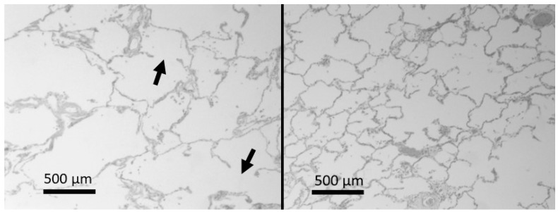
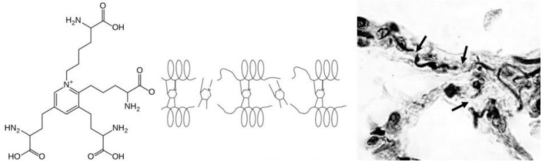
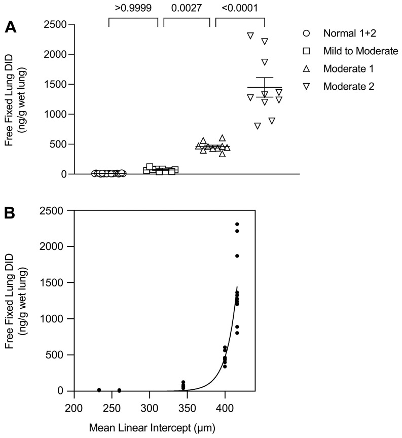
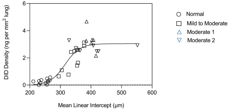
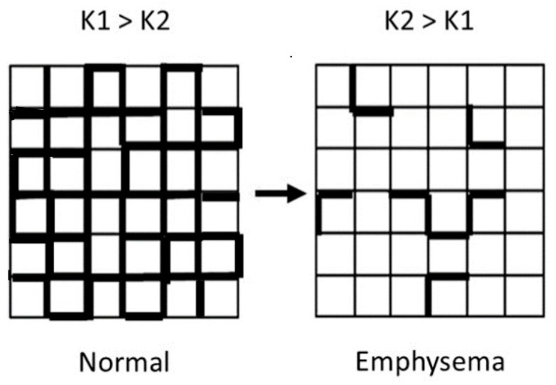
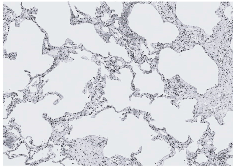
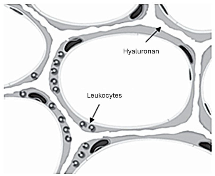
Pulmonary emphysema involves progressive destruction of alveolar walls, leading to enlarged air spaces and impaired gas exchange. While the precise mechanisms responsible for these changes remain unclear, there is growing evidence that the extracellular matrix plays a critical role in the process. An essential feature of pulmonary emphysema is damage to the elastic fiber network surrounding the airspaces, which stores the energy needed to expel air from the lungs. The degradation of these fibers disrupts the mechanical forces involved in respiration, resulting in distension and rupture of alveolar walls. While the initial repair process mainly consists of elastin degradation and resynthesis, continued alveolar wall injury may be associated with increased collagen deposition, resulting in a mixed pattern of emphysema and interstitial fibrosis. Due to the critical role of elastic fiber injury in pulmonary emphysema, preventing damage to this matrix component has emerged as a potential therapeutic strategy. One treatment approach involves the intratracheal administration of hyaluronan, a polysaccharide that prevents elastin breakdown by binding to lung elastic fibers. In clinical trials, inhalation of aerosolized HA decreased elastic fiber injury, as measured by the release of the elastin-specific cross-linking amino acids, desmosine, and isodesmosine. By protecting elastic fibers from enzymatic and oxidative damage, aerosolized HA could alter the natural history of pulmonary emphysema, thereby reducing the risk of respiratory failure.

Keywords: collagen; elastin; extracellular matrix; hyaluronan; pulmonary emphysema.


Conflict of interest statement

Jerome Cantor is an inventor on USPTO patent number 10933084 entitled “Compositions and Methods for Treating Elastic Fiber Breakdown”.

Figures

Figure 1
Figure 1
(Left) Photomicrograph of a lung with emphysema showing airspace enlargement and alveolar wall rupture (arrows). (Right) Photomicrograph of normal lung for comparison. Hematoxylin and eosin. Reprinted with permission [17].
Figure 2
Figure 2
(Left) Desmosine is formed by condensation of lysyl residues on adjacent elastin peptides. (Center) Loss of elastin cross-links results in unraveling and fragmentation of elastic fibers. (Right) Photomicrograph of fragmented elastic fibers (arrows), reprinted with permission [17].
Figure 3
Figure 3
(A) Graph showing the release of free DID (not peptide-bound) from normal lungs and those with mild to moderate or moderate emphysema (as indicated by legend symbols). (B) Graph showing the relationship between free lung DID and alveolar diameter. A phase transition involving greatly increased loss of elastin cross-links occurs around 400 µm. (A,B) reprinted with permission [17].
Figure 4
Figure 4
Graph showing the relationship between DID density and alveolar diameter in normal lungs and those with mild to moderate or moderate emphysema (as indicated by legend symbols). A phase transition consisting of a marked increase in cross-link density occurs between 300 and 400 µm. Reprinted with permission [17].
Figure 5
Figure 5
The progression of pulmonary emphysema involves a transition from intact (strong) to fragmented (weak) elastic fibers. The mechanism responsible for this change involves enzymatic and oxidative degradation of the fibers, which increases the mechanical strain on alveolar walls, resulting in their distention and rupture [32].
Figure 6
Figure 6
Photomicrograph of a human lung with CPFE, showing a mixed pattern of airspace enlargement and interstitial fibrosis.
Figure 7
Figure 7
Cartoon illustrating the binding of intratracheally instilled HA to alveolar wall elastic fibers, which protects them from damage due to leukocyte elastases [12].

Similar articles

Cited by

References

    1. Rabe K.F., Watz H. Chronic obstructive pulmonary disease. Lancet. 2017;389:1931–1940. doi: 10.1016/S0140-6736(17)31222-9. - DOI - PubMed
    1. Churg A., Wright J.L. Proteases and emphysema. Curr. Mol. Med. 2006;6:289–308. doi: 10.1097/01.mcp.0000149592.51761.e3. - DOI - PubMed
    1. Burgess J.K., Mauad T., Tjin G., Karlsson J.C., Westergren-Thorsson G. The extracellular matrix—The under-recognized element in lung disease? J. Pathol. 2016;240:397–409. doi: 10.1002/path.4808. - DOI - PMC - PubMed
    1. Kulkarni T., O’Reilly P., Antony V.B., Gaggar A., Thannickal V.J. Matrix Remodeling in Pulmonary Fibrosis and Emphysema. Am. J. Respir. Cell Mol. Biol. 2016;54:751–760. doi: 10.1165/rcmb.2015-0166PS. - DOI - PMC - PubMed
    1. White E.S. Lung extracellular matrix and fibroblast function. Ann. Am. Thorac. Soc. 2015;12((Suppl. S1)):S30–S33. doi: 10.1513/AnnalsATS.201406-240MG. - DOI - PMC - PubMed

LinkOut - more resources