. 2024 Oct 3;25(19):10642.
doi: 10.3390/ijms251910642.

Cordyceps militaris Grown on Germinated Rhynchosia nulubilis (GRC) Encapsulated in Chitosan Nanoparticle (GCN) Suppresses Particulate Matter (PM)-Induced Lung Inflammation in Mice

Affiliations

Cordyceps militaris Grown on Germinated Rhynchosia nulubilis (GRC) Encapsulated in Chitosan Nanoparticle (GCN) Suppresses Particulate Matter (PM)-Induced Lung Inflammation in Mice

Byung-Jin Park et al. Int J Mol Sci. .

Abstract

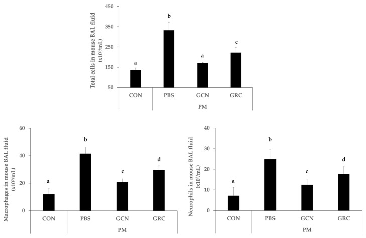
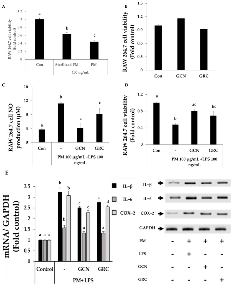
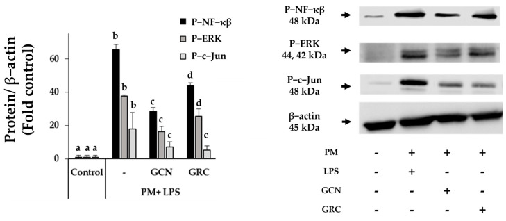
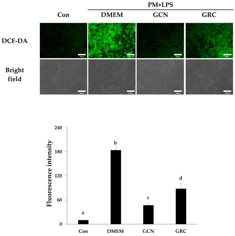
Cordyceps militaris grown on germinated Rhynchosia nulubilis (GRC) exerts various biological effects, including anti-allergic, anti-inflammatory, and immune-regulatory effects. In this study, we investigated the anti-inflammatory effects of GRC encapsulated in chitosan nanoparticles (CN) against particulate matter (PM)-induced lung inflammation. Optimal CN (CN6) (CHI: TPP w/w ratio of 4:1; TPP pH 2) exhibited a zeta potential of +22.77 mV, suitable for GRC encapsulation. At different GRC concentrations, higher levels (60 and 120 mg/mL) led to increased negative zeta potential, enhancing stability. The optimal GRC concentration for maximum entrapment (31.4 ± 1.35%) and loading efficiency (7.6 ± 0.33%) of GRC encapsulated in CN (GCN) was 8 mg/mL with a diameter of 146.1 ± 54 nm and zeta potential of +30.68. In vivo studies revealed that administering 300 mg/kg of GCN significantly decreased the infiltration of macrophages and T cells in the lung tissues of PM-treated mice, as shown by immunohistochemical analysis of CD4 and F4/80 markers. Additionally, GCN ameliorated PM-induced lung tissue damage, inflammatory cell infiltration, and alveolar septal hypertrophy. GCN also decreased total cells and neutrophils, showing notable anti-inflammatory effects in the bronchoalveolar lavage fluid (BALF) from PM-exposed mice, compared to GRC. Next the anti-inflammatory properties of GCN were further explored in PM- and LPS-exposed RAW264.7 cells; it significantly reduced PM- and LPS-induced cell death, NO production, and levels of inflammatory cytokine mRNAs (IL-1β, IL-6, and COX-2). GCN also suppressed NF-κB/MAPK signaling pathways by reducing levels of p-NF-κB, p-ERK, and p-c-Jun proteins, indicating its potential in managing PM-related inflammatory lung disease. Furthermore, GCN significantly reduced PM- and LPS-induced ROS production. The enhanced bioavailability of GRC components was demonstrated by an increase in fluorescence intensity in the intestinal absorption study using FITC-GCN. Our data indicated that GCN exhibited enhanced bioavailability and potent anti-inflammatory and antioxidant effects in cells and in vivo, making it a promising candidate for mitigating PM-induced lung inflammation and oxidative stress.

Keywords: C. militaris grown on germinated R. nulubilis; Chitosan nanoparticles; particulate matter anti-inflammatory activity; respiratory disease.


Conflict of interest statement

Author Kyu-Ree Dhong was employed by the company Magicbullettherapeutics Inc. The remaining authors declare that the research was conducted in the absence of any commercial or financial relationships that could be construed as a potential conflict of interest.

Figures

Figure 1
Figure 1
Optimal GRC encapsulated in chitosan nanoparticles (GCN) synthesis. (A) Particle sizes, concentrations, and zeta potentials of chitosan (CHI) nanoparticles (CN) with different CHI ratios at pH 9.8 and 2.and (B) Representative NTA images of GCN (2, 4, and 8 mg/mL) and particle size distribution and zeta potential results of GCN at various GRC concentrations (2, 4, 8, 30, 60, and 120 mg/mL).
Figure 2
Figure 2
GCN mitigates macrophage and T-helper-cells infiltration and reduces inflammatory gene expression in lung tissue of PM-stimulated mice. (A) Representative images of CD4 and F4/80 immunohistochemical staining (scale bar: 50 µm, magnification: 200×). and (B) lung tissue of control, PM, PM + GCN and PM + GRC-treated mice stained with hematoxylin and eosin (H&E) (scale bar: 50 µm, 200× magnification) and (C) the total number of cells in the lung tissue section. The experiments were repeated three times (n > 3) and results are presented as mean ± SD. a–c Bars with different letters differ significantly at p < 0.05 by Tukey HSD test. PM (particulate matter), GCN (GRC encapsulated in chitosan nanoparticles). Control = mouse treated with PBS; negative control group = mouse treated with particulate matter and PBS; GCN = mouse treated with particulate matter and (GCN 300 mg/kg/day); GRC = mouse treated with particulate matter and GRC (300 mg/kg/day).
Figure 3
Figure 3
GCN reduced the number of infiltrated total cells in bronchoalveolar lavage fluid (BALF) of mice with PM-induced airway inflammation. Total number of cells in BALF of 6-week PM-exposed mice by group. The experiments were repeated three times (n > 3) and results are presented as mean ± SD. a–d Bars with different letters differ significantly at p < 0.05 by Tukey HSD test. PM (particulate matter), GCN (GRC encapsulated in chitosan nanoparticles). Control = mouse treated with PBS; negative control group = mouse treated with particulate matter and PBS; GCN = mouse treated with particulate matter and (GCN 300 mg/kg/day); GRC = mouse treated with particulate matter and GRC (300 mg/kg/day).
Figure 4
Figure 4
GCN reduces inflammatory gene expression in lung tissue of PM-exposed mice. The level of TNF−α, IL−5, IL−1β, and INF-γ mRNAs expression were measured. The experiments were repeated three times (n > 3) and results are presented as mean ± SD. a−d Bars with different letters differ significantly at p < 0.05 by Tukey HSD test. PM (particulate matter), GCN (GRC encapsulated in chitosan nanoparticles). Control = mouse treated with PBS; negative control group = mouse treated with particulate matter and PBS; GCN = mouse treated with particulate matter and GCN (300 mg/kg/day); GRC = mouse treated with particulate matter and GRC (300 mg/kg/day).
Figure 5
Figure 5
GCN inhibits particulate matter (PM)- and lipopolysaccharide (LPS)-induced RAW264.7 cell inflammatory mediator expression. (A) The viability of RAW264.7 cells exposed to wet heat sterilized PM and unsterilized PM (100 μg/mL) was determined by the cell counting kit-8 (CCK-8) assay. (B) Viability of RAW264.7 cells after GCN and GRC (200 μg/mL) treatment was measured using a CCK-8 assay. (C) Nitric Oxide (NO) production in PM (100 μg/mL)- and LPS (100 ng/mL)-treated RAW 264.7 in the presence or the absence of GRC or GCN (200 μg/mL). (D) Cell viability of GRC and GCN (200 μg/mL) treatment in PM (100 μg/mL) and LPS (100 ng/mL) stimulated RAW264.7. (E) Effects of GCN and GRC (200 μg/mL) on the levels of inflammatory cytokine mRNAs in RAW 264.7 cells treated with PM (100 μg/mL) and LPS (100 ng/mL). The experiments were repeated three times and results are presented as mean ± SD. a–d Bars with different letters differ significantly at p < 0.05 by Tukey HSD test.
Figure 6
Figure 6
GCN attenuates PM- and LPS-induced inflammatory responses by suppressing the activation of NF-κB and MAPK signaling pathways. RAW 264.7 cells were pre-treated with 200 μg/mL GRC or GCN for 1 h, followed by treatment with PM (100 µg/mL) and LPS (100 ng/mL) for 24 h. Whole-cell lysates were processed for western blot analysis and probed with indicated antibodies. Phosphorylated NF-κB (p-NF-κB), ERK (p-ERK), and c-Jun (p-c-Jun) protein expression levels in RAW 264.7 cells were detected using western blotting. The experiments were repeated three times and results are presented as mean ± SD. a–d Bars with different letters differ significantly at p < 0.05 by Tukey HSD test.
Figure 7
Figure 7
Antioxidant effects of GCN in particulate matter (PM)- and lipopolysaccharide (LPS)-treated RAW264.7 cells. RAW264.7 cells were treated with GRC or GCN (200 µg/mL) for 1 h followed by treatment with PM (100 µg/mL) and LPS (100 ng/mL) for 3 h. Intracellular ROS were detected by fluorescence microscopy (Nikon Eclipse Ti microscope, Point Grey Research, Richmond, BC, Canada) after 2’,7’-dichlorodihydrofluorescein diacetate (DCF-DA) staining, using Metamorph software version 7.8 (Universal Imaging, West Chester, PA, USA; magnification = 200×; scale bar = 100 µm). Relative fluorescence intensities were analyzed using ImageJ software v1.54k. The experiments were repeated three times and results are presented as mean ± SD. a–d Bars with different letters differ significantly at p < 0.05 by Tukey HSD test.
Figure 8
Figure 8
Analysis of GRC encapsulation in chitosan nanoparticles (GCN) mucoadhesion in vivo. Confocal images of excised small intestinal after oral administration of FITC-GRC or FITC-GCN in mice. The dose was equivalent to 300 mg/kg FTIC-GRC or FITC-GCN, and small intestinal sections were taken 3 h after dosage (Scale bar: 100 μm, Magnification: ×100). Relative fluorescence intensities were analyzed using ImageJ software v1.54k. The experiments were repeated three times and results are presented as mean ± SD. a–c Bars with different letters differ significantly at p < 0.05 by Tukey HSD test. PM (particulate matter), GCN (GRC encapsulated in chitosan nanoparticles).
Figure 9
Figure 9
Particulate matter (PM) nebulize schedule. Mice were exposed to PM via inhalation for 4 weeks, followed by an additional week of intranasal instillation (5 days/week, n = 6 per group). Mice were treated by oral gavage with 100 μL of PBS or sample (300 mg/kg) daily for 6 weeks (5 days/week) and tissues were collected 2 days later for analysis.

Similar articles

Cited by

References

    1. Schraufnagel D.E. The health effects of ultrafine particles. Exp. Mol. Med. 2020;52:311–317. doi: 10.1038/s12276-020-0403-3. - DOI - PMC - PubMed
    1. Wang T., Wang L., Moreno-Vinasco L., Lang G.D., Siegler J.H., Mathew B., Usatyuk P.V., Samet J.M., Geyh A.S., Breysse P.N. Particulate matter air pollution disrupts endothelial cell barrier via calpain-mediated tight junction protein degradation. Part. Fibre Toxicol. 2012;9:35. doi: 10.1186/1743-8977-9-35. - DOI - PMC - PubMed
    1. Dai X., Liu H., Chen D., Zhang J. Association between ambient particulate matter concentrations and hospitalization for ischemic heart disease (I20-I25, ICD-10) in China: A multicity case-crossover study. Atmos. Environ. 2018;186:129–135. doi: 10.1016/j.atmosenv.2018.05.033. - DOI
    1. Kim K.-H., Kabir E., Kabir S. A review on the human health impact of airborne particulate matter. Environ. Int. 2015;74:136–143. doi: 10.1016/j.envint.2014.10.005. - DOI - PubMed
    1. Fiordelisi A., Piscitelli P., Trimarco B., Coscioni E., Iaccarino G., Sorriento D. The mechanisms of air pollution and particulate matter in cardiovascular diseases. Heart Fail. Rev. 2017;22:337–347. doi: 10.1007/s10741-017-9606-7. - DOI - PubMed