. 2024 Oct 3;25(19):10649.
doi: 10.3390/ijms251910649.

Exploring Lysophosphatidylcholine as a Biomarker in Ischemic Stroke: The Plasma-Brain Disjunction

Affiliations

Exploring Lysophosphatidylcholine as a Biomarker in Ischemic Stroke: The Plasma-Brain Disjunction

Justin Turpin et al. Int J Mol Sci. .

Abstract

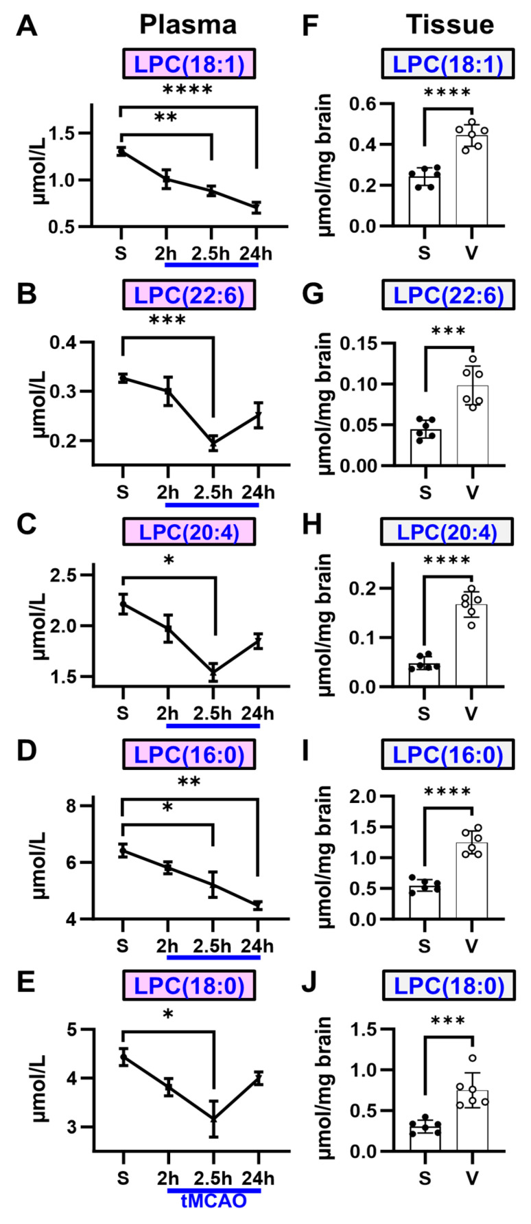
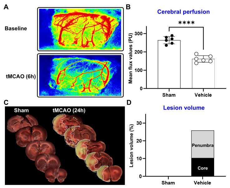
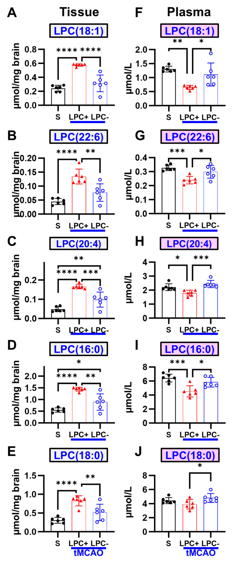
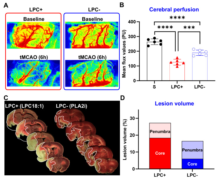
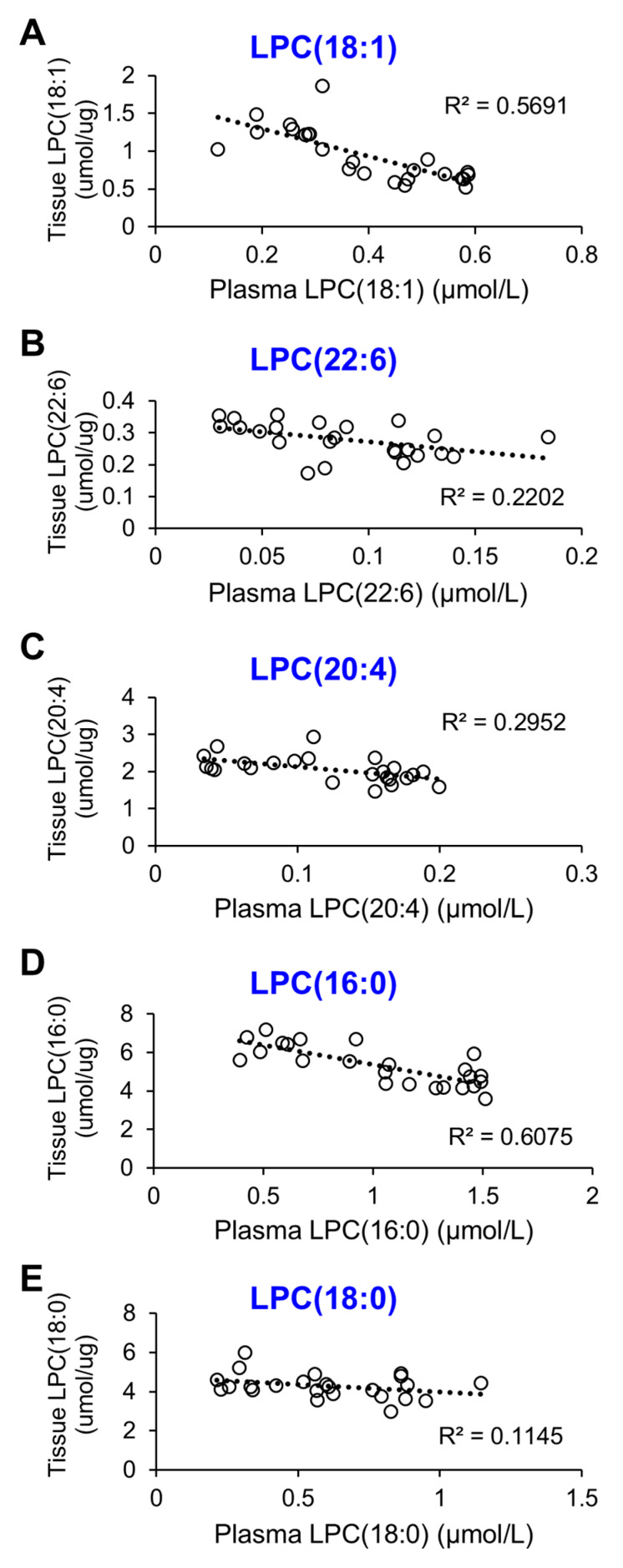
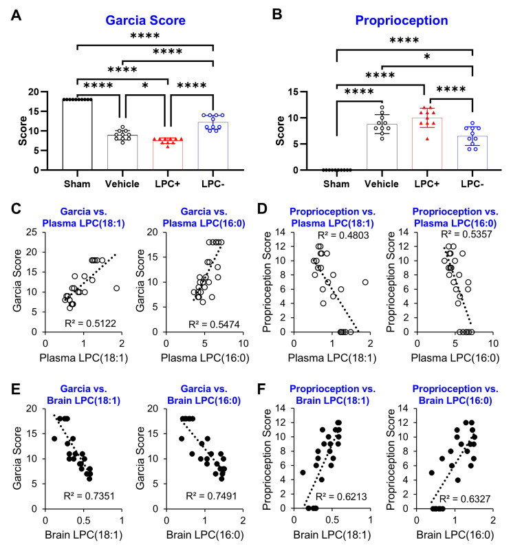
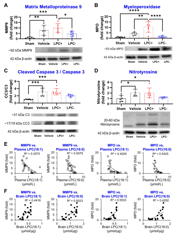
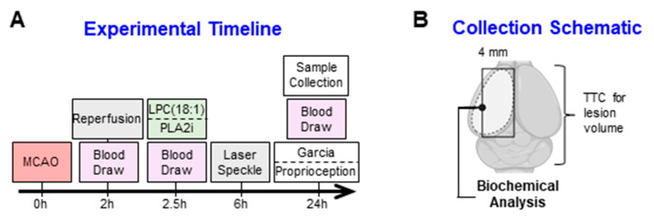
Lipids and their bioactive metabolites, notably lysophosphatidylcholine (LPC), are increasingly important in ischemic stroke research. Reduced plasma LPC levels have been linked to stroke occurrence and poor outcomes, positioning LPC as a potential prognostic or diagnostic marker. Nonetheless, the connection between plasma LPC levels and stroke severity remains unclear. This study aimed to elucidate this relationship by examining plasma LPC levels in conjunction with brain LPC levels to provide a deeper understanding of the underlying mechanisms. Adult male Sprague-Dawley rats underwent transient middle cerebral artery occlusion and were randomly assigned to different groups (sham-operated, vehicle, LPC supplementation, or LPC inhibition). We measured multiple LPC species in the plasma and brain, alongside assessing sensorimotor dysfunction, cerebral perfusion, lesion volume, and markers of BBB damage, inflammation, apoptosis, and oxidative stress. Among five LPC species, plasma LPC(16:0) and LPC(18:1) showed strong correlations with sensorimotor dysfunction, lesion severity, and mechanistic biomarkers in the rat stroke model. Despite notable discrepancies between plasma and brain LPC levels, both were strongly linked to functional outcomes and mechanistic biomarkers, suggesting that LPC's prognostic value is retained extracranially. This study advances the understanding of LPC as a blood marker in ischemic stroke and highlights directions for future research to further elucidate its association with stroke severity, particularly through investigations in more clinically representative models.

Keywords: LPC(18:1); biomarker; ischemic stroke; lipid; lysophosphatidylcholine; prognostic marker; transient middle cerebral artery occlusion.


Conflict of interest statement

The authors declare no conflicts of interest.

Figures

Figure 1
Figure 1
Plasma LPC levels decrease, while brain LPC levels increase following ischemic stroke. (AE) Plasma concentrations of all five LPC species progressively decline following MCAO and reperfusion. Notably, LPC(18:1) and LPC(16:0) show a marked decrease between 2 and 24 h, with significant differences from sham levels observed at the 24 h mark. (FJ) Conversely, all five LPC species exhibit a significant increase in brain tissue at 24 h post-MCAO. (n = 6, * p < 0.05, ** p < 0.01, *** p < 0.001, **** p < 0.0001).
Figure 2
Figure 2
Ischemic stroke leads to reduced cerebral perfusion and induces substantial lesion formation during the acute phase. (A,B) At 6 h post-MCAO induction, cerebral perfusion is significantly diminished. (C,D) By 24 h post-MCAO, a significant lesion develops on the ipsilateral side, encompassing a large portion of core tissue (n = 6, **** p < 0.0001).
Figure 3
Figure 3
LPC supplementation and inhibition of LPC formation influence both plasma and brain LPC levels following ischemic stroke. (AE) Brain levels of all LPC species are elevated above vehicle levels with LPC(18:1) supplementation at 24 h, whereas the inhibition of LPC formation leads to a reduction in all species. (FJ) In plasma, LPC levels do not fall below vehicle levels at 24 h with supplementation, while inhibition results in increased plasma LPC levels. (n = 6, * p < 0.05, ** p < 0.01, *** p < 0.001, **** p < 0.0001, black circles = sham, red triangles = LPC+, blue open circles = LPC−).
Figure 4
Figure 4
LPC supplementation and inhibition of LPC formation modulate cerebral perfusion and lesion volume following ischemic stroke. (A,B) LPC supplementation exacerbates the reduction in cerebral perfusion at 6 h, while the inhibition of LPC formation mitigates the perfusion decrease. (C,D) At 24 h post-MCAO, LPC supplementation results in an increase in the core lesion volume without altering the total lesion volume compared to the vehicle group, while the inhibition of LPC formation reduces both total and core lesion volumes. (n = 6, *** p < 0.001, **** p < 0.0001, black circles = sham, red triangles = LPC+, blue open circles = LPC−).
Figure 5
Figure 5
Plasma LPC levels display a negative correlation with brain LPC levels across all five species, with an increase in brain LPC corresponding to a decrease in plasma LPC. Notably, LPC(18:1) and LPC(16:0) are the only species that exhibit a strong correlation across all LPC species. (A) LPC(18:1). (B) LPC(22:6). (C) LPC(20:4). (D) LPC(16:0). (E) LPC(18:0).
Figure 6
Figure 6
Plasma LPC levels are strongly associated with sensorimotor dysfunction. (A,B) At 24 h post-MCAO induction, rats show significant sensorimotor and proprioceptive impairments, which are exacerbated by LPC(18:1) supplementation and mitigated by LPC formation inhibition. (C,D) Plasma LPC(18:1) and LPC(16:0) levels demonstrate moderate correlations with sensorimotor outcomes. (E,F) Brain LPC(18:1) and LPC(16:0) levels show strong correlations with sensorimotor outcomes. (n = 6, * p < 0.05, **** p < 0.0001, Bar graphs: black circles = sham, red triangles = LPC+, blue open circles = LPC−, Correlation graphs: black circles = Plasma LPC, open circles = Brain LPC).
Figure 7
Figure 7
Plasma LPC levels in ischemic stroke are correlated with established prognostic markers. (AD) At 24 h post-MCAO induction, brain levels of MMP-9, MPO, CC3/C3, and Nitrotyrosine significantly increase, reflecting BBB damage, neutrophil infiltration, apoptosis, and oxidative stress, respectively. (E,F) Both plasma and brain levels of LPC(18:1) and LPC(16:0) display moderate correlations with MMP-9 and MPO expression in the brain. (n = 6, * p < 0.05, ** p < 0.01, *** p < 0.001, **** p < 0.0001, Bar graphs: black circles = sham, red triangles = LPC+, blue open circles = LPC−, Correlation graphs: black circles = Plasma LPC, open circles = Brain LPC).
Figure 8
Figure 8
Conceptual diagram of the experimental protocol. (A) The timeline illustrates the MCAO and drug-administration procedures. Animals were subjected to 2 h of ischemia followed by reperfusion. At 2.5 h post-MCAO induction, animals received either LPC(18:1) or a PLA2 inhibitor. Cerebral perfusion was assessed 6 h after ischemia, and sensorimotor function was evaluated immediately before sample collection at 24 h post-MCAO. (B) Samples for biochemical analyses were collected from the ipsilateral penumbra, with lesion analysis performed using TTC staining.

Similar articles

References

    1. Herpich F., Rincon F. Management of Acute Ischemic Stroke. Crit. Care Med. 2020;48:1654. doi: 10.1097/CCM.0000000000004597. - DOI - PMC - PubMed
    1. Kwakkel G., Veerbeek J.M., Harmeling-van der Wel B.C., van Wegen E., Kollen B.J., on behalf of the Early Prediction of functional Outcome after Stroke (EPOS) Investigators Diagnostic Accuracy of the Barthel Index for Measuring Activities of Daily Living Outcome after Ischemic Hemispheric Stroke. Stroke. 2011;42:342–346. doi: 10.1161/STROKEAHA.110.599035. - DOI - PubMed
    1. Fahey M., Crayton E., Wolfe C., Douiri A. Clinical prediction models for mortality and functional outcome following ischemic stroke: A Syst. review and meta-analysis. PLoS ONE. 2018;13:e0185402. doi: 10.1371/journal.pone.0185402. - DOI - PMC - PubMed
    1. Montellano F.A., Ungethüm K., Ramiro L., Nacu A., Hellwig S., Fluri F., Whiteley W.N., Bustamante A., Montaner J., Heuschmann P.U. Role of Blood-Based Biomarkers in Ischemic Stroke Prognosis. Stroke. 2021;52:543–551. doi: 10.1161/STROKEAHA.120.029232. - DOI - PubMed
    1. Whiteley W., Chong W.L., Sengupta A., Sandercock P. Blood Markers for the Prognosis of Ischemic Stroke. Stroke. 2009;40:e380–e389. doi: 10.1161/STROKEAHA.108.528752. - DOI - PubMed

LinkOut - more resources