. 2024 Oct 3;25(19):10679.
doi: 10.3390/ijms251910679.

Utilizing Adenovirus Knob Proteins as Carriers in Cancer Gene Therapy Amidst the Presence of Anti-Knob Antibodies

Affiliations

Utilizing Adenovirus Knob Proteins as Carriers in Cancer Gene Therapy Amidst the Presence of Anti-Knob Antibodies

Naoya Koizumi et al. Int J Mol Sci. .

Abstract

Numerous gene therapy drugs for cancer have received global approval, yet their efficacy against solid tumors remains inadequate. Our previous research indicated that the fiber protein, a component of the adenovirus capsid, can propagate from infected cells to neighboring cells that express the adenovirus receptor. We hypothesize that merging this fiber protein with an anti-cancer protein could enable the anti-cancer protein to disseminate around the transfected cells, presenting a novel approach to cancer gene therapy. In our study, we discovered that the knob region of the adenovirus type 5 fiber protein is the smallest unit capable of spreading to adjacent cells in a receptor-specific manner. We also showed that the recombinant knob protein infiltrates cells after dispersing to surrounding cells. To assess the potential of the knob protein to augment gene therapy for solid tumors in mice, we expressed a fusion gene of the A subunit of cytotoxic cholera toxin and the knob region in mouse tumors. We found that this fusion protein only inhibited tumor growth in receptor-expressing mouse melanomas, and this inhibitory effect persisted even in mice with anti-knob antibodies. Our study's findings propose a novel cancer gene therapy strategy that enhances therapeutic effects by specifically delivering therapeutic proteins, expressed from in vivo administered genes, to target molecules. This outcome offers a fresh perspective on gene therapy for solid cancers, and we anticipate that knob proteins will serve as a platform for this method.

Keywords: cancer; gene therapy; knob protein; neutralizing antibodies; targeting.


Conflict of interest statement

The authors declare that there are no conflicts of interest.

Figures
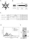
Figure 1
Figure 1
Analysis of adenovirus knob region distribution on 293T Cells. (A,B) Schematic representation of adenovirus (Ad) and Ad fiber protein. (C) Several Ad capsid proteins expressed via plasmid. (D) Flow-cytometry analysis of Ad capsid proteins in 293T cells transfected with plasmids. Twenty-four hours post-transfection, cells were harvested using 2 mM EDTA/PBS and stained with anti-DDDDK monoclonal antibody. (E) Western blot analysis of Ad capsid proteins in 293T cells transfected with plasmids. Twenty-four hours post-transfection, cells were harvested using 2 mM EDTA/PBS and disrupted via ultrasonication. After centrifugation to collect the supernatant, 10 mg of the supernatant was separated on a 15% SDS-polyacrylamide gel, and the Ad capsid proteins were analyzed by Western blotting using an anti-DDDDK monoclonal antibody as outlined in Section 4.
Figure 2
Figure 2
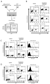
CAR-specific cell-to-cell spread of knob protein (A) Schematic representation of experimental methods. (BD) Flow-cytometry analysis of type 5 Ad knob protein (Ad5knob) on various co-cultured GFP-expressing cells. Twenty-four hours post-transfection of 293T cells, cells were harvested using 2 mM EDTA/PBS and co-cultured with either CAR-positive or CAR-negative cells (B), CAR-knockdown 293T cells (C), or CAR-expressed SF293 cells (D). After 24 h of co-culture, cells were harvested using 2 mM EDTA/PBS and analyzed by flow-cytometry using an anti-DDDDK monoclonal antibody as outlined in Section 4. Data are expressed as mean ± S.D. (n = 3).
Figure 3
Figure 3
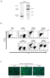
Investigation of cell-to-cell spread of recombinant knob protein. (A) Western blot analysis of recombinant Ad5knob (rAd5knob) protein derived from plasmid-transfected 293T cells. Twenty-four hours post-transfection, cells were harvested using 2 mM EDTA/PBS and disrupted via ultrasonication. Following supernatant collection by centrifugation, rAd5knob was purified using an anti-DDDDK-tag specific gel column. The rAd5knob was then separated on a 15% SDS- or native-polyacrylamide gel and analyzed by Western blotting using an anti-DDDDK monoclonal antibody as outlined in Section 4. (B) Flow-cytometry analysis of rAd5knob on co-cultured 293T-GFP cells. One hour post-co-culture of rAd5knob-binding 293T cells and 293T-GFP cells, cells were analyzed by flow-cytometry using an anti-DDDDK monoclonal antibody as described in Section 4. Mean fluorescence intensity data are expressed as mean (n = 3). (C) Immunohistological staining of rAd5knob in 293T cells. One hour post-Ad5knob binding to 293T cells on ice, cells were cultured for 15 or 30 min at 37 degrees. Cells were stained with anti-DDDDK monoclonal antibody and Hoechst 33342 and observed with a confocal laser microscope.
Figure 4
Figure 4
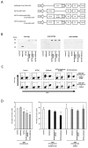
Characteristics of cholera toxin A-subunit and knob fusion protein in vitro. (A) Schematic representation of plasmids expressing Ad5knob fusion proteins. The A subunit of cholera toxin (NCTXA) retains its cytotoxic properties. The efficiency of gene transfer for all plasmids can be confirmed by the quantity of GFP co-expressed with IRES sequences. (B) Western blot analysis of Ad knob fusion proteins in 293T cells transfected with plasmids. The Ad knob fusion proteins were analyzed by Western blotting using an anti-DDDDK monoclonal antibody and an anti-cholera toxin monoclonal antibody as outlined in Section 4. (C) Flow-cytometry analysis of Ad knob fusion proteins on co-cultured cells. Twenty-four hours post-transfection of 293T cells with plasmids, cells were co-cultured with either B16-BL6-hCAR or B16-BL6 cells. After 24 h of co-culture, cells were harvested using 2 mM EDTA/PBS and analyzed by flow-cytometry using an anti-DDDDK monoclonal antibody as described in Section 4. Data are expressed as mean ± S.D. (n = 3). (D) Cell proliferation analysis of NCTXA and Ad knob fusion proteins in co-cultured cells. Twenty-four hours post-transfection of 293T cells with plasmids, cells were cultured alone or co-cultured with either B16-BL6-hCAR or B16-BL6 cells. After 48 h of co-culture, cells were harvested using 2 mM EDTA/PBS, and the number of cells was counted. Statistical analysis was conducted using two-way analysis of variance and the Student–Newman–Keuls post hoc test (* p < 0.05 vs. control).
Figure 5
Figure 5
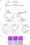
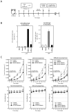
Inhibition of tumor growth in tumor-bearing mice by transfection of genes encoding NCTXA and Ad knob fusion proteins. (A) Timeline of mouse experiments. (B) Inhibition of tumor growth in tumor-bearing mice by intratumoral administration of gene-transfected B16BL6-hCAR cells. Eight days after subcutaneous transplantation of B16BL6-hCAR cells into mice, gene-transfected B16BL6-hCAR cells were intratumorally administered. Subsequently, cells were administered two more times, and tumor diameter and mouse weight were measured. Statistical analysis was conducted using two-way analysis of variance and the Student–Newman–Keuls post hoc test (* p < 0.05 vs. control). (C) Inhibition of tumor growth in tumor-bearing mice by direct gene transfection via intratumoral administration. Seven days after subcutaneous transplantation of B16BL6-hCAR or B16BL6 cells into mice, transfection reagents and plasmids were intratumorally administered. Subsequently, the reagent and plasmids were administered once more, and tumor diameter and weight were measured. Statistical analysis was conducted using two-way analysis of variance and the Student–Newman–Keuls post hoc test (* p < 0.05 vs. NCTXA). (D) Inhibition of tumor growth in tumor-bearing mice by intratumoral administration of gene-transfected B16BL6-hCAR cells. Seven days after subcutaneous transplantation of B16BL6-hCAR cells into mice, gene-transfected B16BL6-hCAR or B16BL6 cells were intratumorally administered. Subsequently, cells were administered once more, and tumor diameter and weight were measured. Statistical analysis was conducted using two-way analysis of variance and the Student–Newman–Keuls post hoc test (* p < 0.05 vs. NCTXA-Ad5knob expressing B16BL6). (E) Preparation of paraffin sections from tumors. Eight days after subcutaneous transplantation of B16BL6-hCAR cells into mice, gene-transfected B16BL6-hCAR cells were intratumorally administered. Two days later, tumor tissue was recovered. Each section was stained with H&E and anti-caspase-3 monoclonal antibody. Data represent means ± SD of three to six mice.
Figure 6
Figure 6
Tumor growth inhibition in Ad knob immunized mice bearing tumors, achieved by transfecting genes encoding NCTXA and Ad knob fusion proteins (A) Timeline for antigen administration in the mouse experiments. (B) Serum antibody levels post-four intraperitoneal administrations of antigens to mice. ELISA was used to measure the antibody levels for Ad5knob or OVA in the serum measured by ELISA. (C) The inhibition of tumor growth in antigen-immunized mice bearing tumors when B16BL6-hCAR cells, transfected with the gene, were administered intratumorally. Eight days post-subcutaneous transplantation of B16BL6-hCAR cells into mice, these gene-transfected cells were administered into the tumor. The tumor diameter and mice weight were then measured. # Mice with high anti-Ad5knob antibody titers. Data represent the means ± SD of three to six mice. Statistical analysis was conducted using two-way analysis of variance and the Student–Newman–Keuls post hoc test (* p < 0.05 vs. control).

Similar articles

References

    1. Hou B., Tang Y., Li W., Zeng Q., Chang D. Efficiency of CAR-T Therapy for Treatment of Solid Tumor in Clinical Trials: A Meta-Analysis. Dis. Markers. 2019;2019:3425291. doi: 10.1155/2019/3425291. - DOI - PMC - PubMed
    1. Wang L., Zhang L., Dunmall L.C., Wang Y.Y., Fan Z., Cheng Z., Wang Y. The dilemmas and possible solutions for CAR-T cell therapy application in solid tumors. Cancer Lett. 2024;591:216871. doi: 10.1016/j.canlet.2024.216871. - DOI - PubMed
    1. Rahman M.M., McFadden G. Oncolytic Viruses: Newest Frontier for Cancer Immunotherapy. Cancers. 2021;13:5452. doi: 10.3390/cancers13215452. - DOI - PMC - PubMed
    1. Boagni D.A., Ravirala D., Zhang S.X. Current Strategies in Engaging Oncolytic Viruses with Antitumor Immunity. Mol. Ther. Oncolytics. 2021;22:98–113. doi: 10.1016/j.omto.2021.05.002. - DOI - PMC - PubMed
    1. Mast T.C., Kierstead L., Gupta S.B., Nikas A.A., Kallas E.G., Novitsky V., Mbewe B., Pitisuttithum P., Schechter M., Vardas E., et al. International Epidemiology of Human Pre-Existing Adenovirus (Ad) Type-5, Type-6, Type-26 and Type-36 Neutralizing Antibodies: Correlates of High Ad5 Titers and Implications for Potential HIV Vaccine Trials. Vaccine. 2010;28:950–957. doi: 10.1016/j.vaccine.2009.10.145. - DOI - PubMed

Grants and funding

LinkOut - more resources