. 2024 Oct 4;25(19):10692.
doi: 10.3390/ijms251910692.

Biocompatible Poly(ε-Caprolactone) Nanocapsules Enhance the Bioavailability, Antibacterial, and Immunomodulatory Activities of Curcumin

Affiliations

Biocompatible Poly(ε-Caprolactone) Nanocapsules Enhance the Bioavailability, Antibacterial, and Immunomodulatory Activities of Curcumin

Floriana D'Angeli et al. Int J Mol Sci. .

Abstract

Curcumin (Cur), the primary curcuminoid found in Curcuma longa L., has garnered significant attention for its potential anti-inflammatory and antibacterial properties. However, its hydrophobic nature significantly limits its bioavailability. Additionally, adipose-derived stem cells (ADSCs) possess immunomodulatory properties, making them useful for treating inflammatory and autoimmune conditions. This study aims to verify the efficacy of poly(ε-caprolactone) nanocapsules (NCs) in improving Cur's bioavailability, antibacterial, and immunomodulatory activities. The Cur-loaded nanocapsules (Cur-NCs) were characterized for their physicochemical properties (particle size, polydispersity index, Zeta potential, and encapsulation efficiency) and stability over time. A digestion test simulated the behavior of Cur-NCs in the gastrointestinal tract. Micellar phase analyses evaluated the Cur-NCs' bioaccessibility. The antibacterial activity of free Cur, NCs, and Cur-NCs against various Gram-positive and Gram-negative strains was determined using the microdilution method. ADSC viability, treated with Cur-NCs and Cur-NCs in the presence or absence of lipopolysaccharide, was analyzed using the 3-[4,5-dimethylthiazol-2-yl]-2,5-diphenyl tetrazolium bromide assay. Additionally, ADSC survival was assessed through the Muse apoptotic assay. The expression of both pro-inflammatory (interleukin-1β and tumor necrosis factor-α) and anti-inflammatory (IL-10 and transforming growth factor-β) cytokines on ADSCs was evaluated by real-time polymerase chain reaction. The results demonstrated high stability post-gastric digestion of Cur-NCs and elevated bioaccessibility of Cur post-intestinal digestion. Moreover, Cur-NCs exhibited antibacterial activity against Escherichia coli without affecting Lactobacillus growth. No significant changes in the viability and survival of ADSCs were observed under the experimental conditions. Finally, Cur-NCs modulated the expression of both pro- and anti-inflammatory cytokines in ADSCs exposed to inflammatory stimuli. Collectively, these findings highlight the potential of Cur-NCs to enhance Cur's bioavailability and therapeutic efficacy, particularly in cell-based treatments for inflammatory diseases and intestinal dysbiosis.

Keywords: ADSCs; LPS; antibacterial activity; bioaccessibility; curcumin; cytokines; immunomodulatory activity; inflammation; poly(ε-caprolactone) nanocapsules; probiotics.


Conflict of interest statement

The authors declare no conflicts of interest.

Figures

Figure 1
Figure 1
Ultraviolet–visible (UV−Vis) spectrum of curcumin-nanocapsules (Cur-NCs) in water. To perform the UV−Vis spectrum, 10 μL of the Cur-NCs nanosuspension was diluted with 3 mL of pure water.
Figure 2
Figure 2
Fluorescence spectra of curcumin-nanocapsules (Cur-NCs) in water and after curcumin (Cur) release in acetonitrile. To perform the fluorescence spectra, 10 μL of the Cur-NCs nanosuspension was diluted with 3 mL of pure water or acetonitrile.
Figure 3
Figure 3
Intensity-weighted distribution of the hydrodynamic diameter (DH) of freshly prepared curcumin-nanocapsules, after 30 days of storage at 25 °C and 40 °C.
Figure 4
Figure 4
Curcumin (Cur) retention percentage in curcumin-nanocapsules (Cur-NCs) after 30 days of storage at 25 °C and 40 °C.
Figure 5
Figure 5
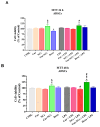
Dose and time-dependent effects of curcumin (Cur), empty nanocapsules (NCs), and curcumin-loaded nanocapsules (Cur-NCs) on primary human adipose-derived stem cells (ADSCs) viability. Cells were grown in a culture medium (CTRL) or exposed to increasing concentrations (0.06–1 µg/mL) of Cur or Cur-NCs or different dilutions of NCs for 24 h (A,A’) and 48 h (B,B’). Results are expressed as a percent of control. The bars represent means ± SD from three independent experiments performed in triplicate (SD = standard deviation). Statistically significant differences, determined by one-way analysis of variance ANOVA and the Tukey post-test, are indicated: * p < 0.05 vs. CTRL at the same incubation time. WS: working standard: diluted solution derived from the stock solution.
Figure 6
Figure 6
Effect of curcumin (Cur), empty nanocapsules (NCs), curcumin-loaded nanocapsules (Cur-NCs), and dexamethasone (Dexa) on primary human adipose-derived stem cells (ADSCs) cell viability, at the steady state, and under inflammatory stimuli. Cells were grown alone (control: CTRL) or in the presence of 0.125 µg/mL of Cur or Cur-NCs, NCs diluted in the 1:8 ratio, 10 nM Dexa, with or without 1 µg/mL lipopolysaccharide (LPS), for 24 h and 48 h. Histograms showed ADSCs cell viability, at 24 h (A) and 48 h (B). Results are expressed as a percent of control. The bars represent means ± SD from three independent experiments performed in triplicate (SD = standard deviation). Statistically significant differences, determined by one-way analysis of variance ANOVA and the Tukey post-test, are indicated: * p < 0.05 vs. CTRL; § p < 0.05 vs. Cur; # p < 0.05 vs. LPS, at the same incubation time.
Figure 7
Figure 7
Evaluation of apoptotic cell death on human adipose-derived stem cells (ADSCs) exposed to curcumin (Cur), empty nanocapsules (NCs), curcumin-loaded nanocapsules (Cur-NCs), dexamethasone (Dexa), and lipopolysaccharide (LPS) in the presence or absence of inflammatory stimuli. A–B Scatter plots of ADSCs grown, for 24 h (A) and 48 h (B), in normal culture medium (control; CTRL) or the presence of 0.125 µg/mL Cur or Cur-NCs, NCs diluted in the 1:8 ratio, 10 nM Dexa, with or without 1 µg/mL LPS. Each plot reports four squares in which cells are distributed based on their staining. (A’,B’) Histograms showed the rate of vital cells (Alive), early apoptotic cells (EA), late apoptotic (LA)/dead cells, and debris for each experimental condition, at 24 h (A’) and 48 h (B’). The bars represent means ± SD of three independent experiments (SD = standard deviation). Statistically significant differences, determined by two-way analysis of variance ANOVA and the Tukey post-test, are indicated: * p < 0.05 vs. CTRL; # p < 0.05 vs. LPS, at the same incubation time.
Figure 7
Figure 7
Evaluation of apoptotic cell death on human adipose-derived stem cells (ADSCs) exposed to curcumin (Cur), empty nanocapsules (NCs), curcumin-loaded nanocapsules (Cur-NCs), dexamethasone (Dexa), and lipopolysaccharide (LPS) in the presence or absence of inflammatory stimuli. A–B Scatter plots of ADSCs grown, for 24 h (A) and 48 h (B), in normal culture medium (control; CTRL) or the presence of 0.125 µg/mL Cur or Cur-NCs, NCs diluted in the 1:8 ratio, 10 nM Dexa, with or without 1 µg/mL LPS. Each plot reports four squares in which cells are distributed based on their staining. (A’,B’) Histograms showed the rate of vital cells (Alive), early apoptotic cells (EA), late apoptotic (LA)/dead cells, and debris for each experimental condition, at 24 h (A’) and 48 h (B’). The bars represent means ± SD of three independent experiments (SD = standard deviation). Statistically significant differences, determined by two-way analysis of variance ANOVA and the Tukey post-test, are indicated: * p < 0.05 vs. CTRL; # p < 0.05 vs. LPS, at the same incubation time.
Figure 8
Figure 8
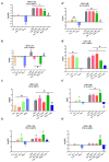
Expression levels of inflammatory cytokines IL-1β, TNF-α, IL-10, and TGF-β on human adipose-derived stem cells (ADSCs) exposed to curcumin (Cur), empty nanocapsules (NCs), curcumin-loaded nanocapsules (Cur-NCs), dexamethasone (Dexa), with or without lipopolysaccharide (LPS), for 24 h and 48 h. ADSCs were cultured in normal culture medium (control; CTRL) or the presence of 0.125 µg/mL Cur or Cur-NCs, NCs diluted in the 1:8 ratio, 10 nM Dexa, with or without 1 µg/mL LPS, for 24 h (AD) and 48 h (A’D’). Histograms showed IL-1β, TNF-α, IL-10, and TGF-β mRNA expression levels on ADSCs grown in our experimental conditions for 24 h (AD) and 48 h (A’D’). The bars represent means ± SD of three independent experiments (SD = standard deviation). Statistically significant differences, determined by one-way analysis of variance ANOVA and the Tukey post-test, are indicated: * p < 0.05 vs. CTRL; # p < 0.05 vs. LPS, at the same incubation time.

Similar articles

References

    1. Chen L., Deng H., Cui H., Fang J., Zuo Z., Deng J., Li Y., Wang X., Zhao L. Inflammatory responses and inflammation-associated diseases in organs. Oncotarget. 2018;9:7204–7218. doi: 10.18632/oncotarget.23208. - DOI - PMC - PubMed
    1. Takeuchi O., Akira S. Pattern recognition receptors and inflammation. Cell. 2010;140:805–820. doi: 10.1016/j.cell.2010.01.022. - DOI - PubMed
    1. Feehan K.T., Gilroy D.W. Is Resolution the End of Inflammation? Trends Mol. Med. 2019;25:198–214. doi: 10.1016/j.molmed.2019.01.006. - DOI - PubMed
    1. Arango Duque G., Descoteaux A. Macrophage cytokines: Involvement in immunity and infectious diseases. Front. Immunol. 2014;5:491. doi: 10.3389/fimmu.2014.00491. - DOI - PMC - PubMed
    1. Striz I., Brabcova E., Kolesar L., Sekerkova A. Cytokine networking of innate immunity cells: A potential target of therapy. Clin. Sci. 2014;126:593–612. doi: 10.1042/CS20130497. - DOI - PubMed

MeSH terms

LinkOut - more resources