Review
. 2024 Oct 6;25(19):10760.
doi: 10.3390/ijms251910760.

Induced Necroptosis and Its Role in Cancer Immunotherapy

Affiliations
Review

Induced Necroptosis and Its Role in Cancer Immunotherapy

Ziyao Zhang et al. Int J Mol Sci. .

Abstract

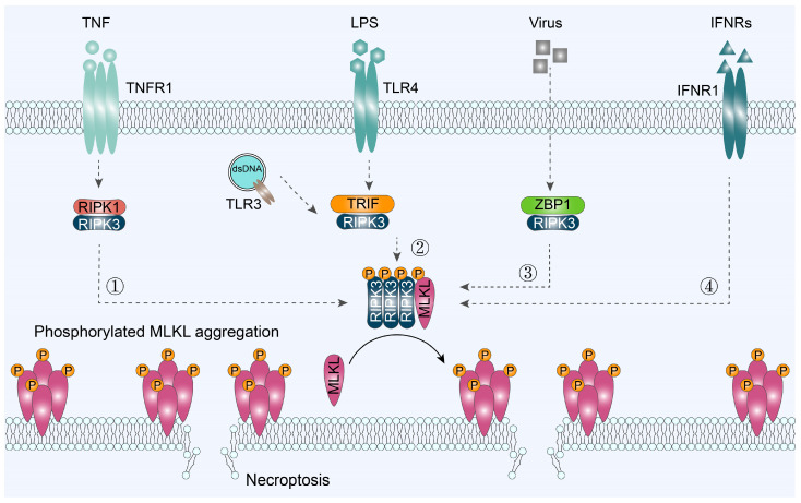
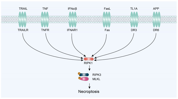
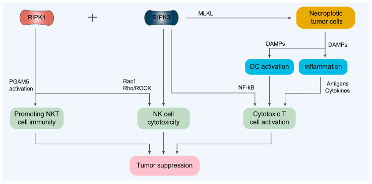
Necroptosis is a type of regulated cell death (RCD) that is triggered by changes in the extracellular or intracellular milieu that are picked up by certain death receptors. Thanks to its potent capacity to induce immunological responses and overcome apoptotic resistance, it has garnered significant attention as a potential cancer treatment. Basic information for the creation of nano-biomedical treatments is provided by studies on the mechanisms underlying tumor necroptosis. Receptor-interacting protein kinase 1 (RIPK1)-RIPK3-mediated necroptosis, Toll-like receptor domain-containing adapter-inducing interferon (IFN)-β (TRIF)-RIPK3-mediated necroptosis, Z-DNA-binding protein 1 (ZBP1)-RIPK3-mediated necroptosis, and IFNR-mediated necroptosis are the four signaling pathways that collectively account for triggered necroptosis in this review. Necroptosis has garnered significant interest as a possible cancer treatment strategy because, in contrast to apoptosis, it elicits immunological responses that are relevant to therapy. Thus, a thorough discussion is held on the connections between tumor cell necroptosis and the immune environment, cancer immunosurveillance, and cells such as dendritic cells (DCs), cytotoxic T cells, natural killer (NK) cells, natural killer T (NKT) cells, and their respective cytokines. Lastly, a summary of the most recent nanomedicines that cause necroptosis in order to cause immunogenic cell death is provided in order to emphasize their promise for cancer immunotherapy.

Keywords: DAMPs; cancer immunotherapy; immune microenvironment; immunogenic cell death; necroptosis.


Conflict of interest statement

The authors declare no conflicts of interest.

Figures

Figure 1
Figure 1
The timeline records the development of necroptosis for cancer treatment. RIPK1, receptor-interacting protein kinase 1; RIPK3, receptor-interacting protein kinase 3; MLKL, mixed-lineage kinase domain-like protein; ZBP1, Z-DNA binding protein 1; TRIF (also called TICAM-1), TIR domain-containing adapter-inducing interferon-β; TLR3, Toll-like receptor 3; TLR4, Toll-like receptor 4; ADAR1, adenosine deaminase acting on RNA enzyme-1.
Figure 2
Figure 2
Signaling pathways to trigger cell necroptosis. The induced necroptosis is summarized into four signaling pathways: ① RIPK1/RIPK3/MLKL-, ② TRIF/RIPK3/MLKL-, ③ ZBP1/RPK3/LKKL-, and ④ IFNR/MLKL-mediated necroptosis. TNF, tumor necrosis factor; TNFR1, tumor necrosis factor receptor 1; LPS, lipopolysaccharide; TLR3/4, Toll-like receptors 3 and 4; IFNRs, type I/II interferon receptors; IFNR1, interferon alpha and beta receptor subunit 1.
Figure 3
Figure 3
Signaling pathways to trigger cell necroptosis via RIPK1/RIPK3/MLKL axis. TRAIL, TNF-related apoptosis-inducing ligand; TNF, tumor necrosis factor; IFNα/β, interferon α/β; FasL, Fas ligand; TL1A, tumor necrosis factor-like cytokine 1A; APP, amyloid precursor protein.
Figure 4
Figure 4
The immune microenvironment associated with tumor necroptosis. VEGF, vascular endothelial growth factor; FGFs, fibroblast growth factors; DAMPs, damage-associated molecular patterns.
Figure 5
Figure 5
The relationship between necroptosis and cancer immunosurveillance. NF-κB, nuclear factor kappa-B; NKT cell, natural killer T cell.

Similar articles

Cited by

References

    1. Yan J., Wan P., Choksi S., Liu Z.-G. Necroptosis and Tumor Progression. Trends Cancer. 2022;8:21–27. doi: 10.1016/j.trecan.2021.09.003. - DOI - PMC - PubMed
    1. Bertheloot D., Latz E., Franklin B.S. Necroptosis, Pyroptosis and Apoptosis: An Intricate Game of Cell Death. Cell Mol. Immunol. 2021;18:1106–1121. doi: 10.1038/s41423-020-00630-3. - DOI - PMC - PubMed
    1. Lin J., Kumari S., Kim C., Van T.-M., Wachsmuth L., Polykratis A., Pasparakis M. RIPK1 Counteracts ZBP1-Mediated Necroptosis to Inhibit Inflammation. Nature. 2016;540:124–128. doi: 10.1038/nature20558. - DOI - PMC - PubMed
    1. Someda M., Kuroki S., Miyachi H., Tachibana M., Yonehara S. Caspase-8, Receptor-Interacting Protein Kinase 1 (RIPK1), and RIPK3 Regulate Retinoic Acid-Induced Cell Differentiation and Necroptosis. Cell Death Differ. 2020;27:1539–1553. doi: 10.1038/s41418-019-0434-2. - DOI - PMC - PubMed
    1. Rodriguez D.A., Quarato G., Liedmann S., Tummers B., Zhang T., Guy C., Crawford J.C., Palacios G., Pelletier S., Kalkavan H., et al. Caspase-8 and FADD Prevent Spontaneous ZBP1 Expression and Necroptosis. Proc. Natl. Acad. Sci. USA. 2022;119:e2207240119. doi: 10.1073/pnas.2207240119. - DOI - PMC - PubMed

Substances