. 2024 Oct 9;25(19):10854.
doi: 10.3390/ijms251910854.

Chemokine Receptor N-Terminus Charge Dictates Reliance on Post-Translational Modifications for Effective Ligand Capture and Following Boosting by Defense Peptides

Affiliations

Chemokine Receptor N-Terminus Charge Dictates Reliance on Post-Translational Modifications for Effective Ligand Capture and Following Boosting by Defense Peptides

Ting Xu et al. Int J Mol Sci. .

Abstract

The chemokine receptors CCR1 and CCR5 display overlapping expression patterns and ligand dependency. Here we find that ligand activation of CCR5, not CCR1, is dependent on N-terminal receptor O-glycosylation. Release from O-glycosylation dependency is obtained by increasing CCR5 N-terminus acidity to the level of CCR1. Ligand activation of CCR5, not CCR1, drastically improves in the absence of glycosaminoglycans (GAGs). Ligand activity at both CCR1 and CCR5 is boosted by positively charged/basic peptides shown to interact with acidic chemokine receptor N-termini. We propose that receptors with an inherent low N-terminus acidity rely on post-translational modifications (PTMs) to efficiently compete with acidic entities in the local environment for ligand capture. Although crucial for initial ligand binding, strong electrostatic interactions between the ligand and the receptor N-terminus may counteract following insertion of the ligand into the receptor binding pocket and activation, a process that seems to be aided in the presence of basic peptides. Basic peptides bind to the naked CCR1 N-terminus, not the CCR5 N-terminus, explaining the loss of boosting of ligand-induced signaling via CCR5 in cells incapable of glycosylation.

Keywords: CCR1; CCR5; O-glycosylation; chemokine; electrostatic interaction; glycosaminoglycan; post-translational modification; receptor N-terminus; signaling; tyrosine sulfation.


Conflict of interest statement

B.F.V. and F.C.P. have ownership interests in Protein Foundry, LLC and XLock Bioscience, Inc. M.B.C. was employed by Evaxion Biotech and C.K.G. was employed by Glx Analytix APS. The remaining authors declare that the research was conducted in the absence of any commercial or financial relationships that could be construed as potential conflicts of interest. The funders had no role in the design of the study; in the collection, analyses, or interpretation of data; in the writing of the manuscript; or in the decision to publish the results.

Figures

Figure 1
Figure 1
Basic peptides boost signaling induced by basic chemokines, with no effect on signaling induced by acidic chemokines, at both CCR1 and CCR5. Ligand-induced Gαi signaling via CCR5 and CCR1 in the absence or presence of C21TP81-111 measured in CHO-K1 cells transiently transfected to express the chemokine receptors and the cAMP sensor CAMYEL (EPAC). (A) C21TP81-111 boosts the potency of CCL5 and CCL8 in Gαi signaling but has no effect on CCL3-induced signaling via CCR5. (B) Signaling induced by CCL5 and CCL7, but not CCL3, via CCR1 is boosted in the presence of C21TP81-111. Statistical significances were determined by two-way ANOVA with Sidak’s multiple comparisons test (number of independent experiments, n = 3). ** p ≤ 0.01, * p ≤ 0.05, ns = non-significant.
Figure 2
Figure 2
Apart from the chemokine-derived peptide C21TP81-111, basic peptides in the form of host defense peptides also boost signaling of basic chemokines. Ligand-induced Gαi signaling via CCR1 in the absence or presence of C21TP81-111, hBD2, and histatin1 measured in CHO-K1 cells transfected as in Figure 1. (A) None of the basic peptides affected CCL3-induced signaling via CCR1. Statistical significances were determined by two-way ANOVA with Sidak’s multiple comparisons test (n = 3). (B,C) hBD2 boosts the potency of CCL5 and CCL7 in Gαi signaling via CCR1 to the same extent as C21TP81-111. Histatin1 also boosts the potency of CCL5 and CCL7, but only to a minor degree. ** p ≤ 0.01, * p ≤ 0.05, ns = non-significant.
Figure 3
Figure 3
CCL5 signaling through CCR5 and to a lesser extent CCR1 is improved in the absence of GAGs. Ligand-induced Gαi signaling via CCR5 and CCR1 in the absence or presence of C21TP81-111 in CHO GAG-minus and CHO GAG-plus cells transfected as in Figure 1. (A) Both the potency and the efficacy of CCL5 are significantly increased in GAG-minus cells compared to GAG-plus cells in the absence of C21TP81-111 boosting. Thus, although boosting persists in the GAG-deficient cells, it is limited how much boosting can occur when signaling in the absence of basic peptides is already very potent. (B) In contrast to CCL5 signaling via CCR5, signaling induced by the same ligand via CCR1 is potent in both GAG-deficient and WT CHO cells, although signaling in CHO deficient cells still exceeds that observed in WT CHO cells. Again, since signaling is potent in both GAG-plus and GAG-minus cells, boosting exerted by C21TP81-111although significant, is limited. Statistical significances were determined by two-way ANOVA with Sidak’s multiple comparisons test (n = 3). ** p ≤ 0.01, ns = non-significant.
Figure 4
Figure 4
Chemokine signaling via CCR5, not CCR1, is highly dependent on receptor O-glycosylation. Ligand-induced Gαi signaling via CCR5 and CCR1 in the absence or presence of C21TP81-111 in HEK293 WT, DeltaSia, and sC cells transfected as in Figure 1. (A) Compared to CCL5 signaling in WT cells, both potency and efficacy of CCL5 significantly drop in DeltaSia cells and even more so in sC, while C21TP81-111 boosting seems to persist in all cell lines. CCL3- and CCL8-induced signaling is drastically reduced in DeltaSia and sC cells and boosting of CCL8 disappears. (B) In contrast, CCL5, CCL3, and CCL7 signaling via CCR1 persists in DeltaSia and sC to almost the same high level as observed in WT cells. Boosting of CCL5 and CCL7 signaling also persists. Signaling by CCL3, as expected, is not boosted in the presence of C21TP81-111. Statistical significances were determined by two-way ANOVA with Sidak’s multiple comparisons test (n = 3). ** p ≤ 0.01, * p ≤ 0.05, ns = non-significant.
Figure 5
Figure 5
Receptors are expressed at the same level on the surface of WT, DeltaSia, and sC HEK293. Cell surface expression levels of CCR1 and CCR5 determined using M1-tagged receptors (AC). M1-CCR1 and M1-CCR5 are expressed at the cell surface at similar levels to each other within all three cell lines, HEK293 WT (green columns, A), DeltaSia (blue columns, B), and sC (red columns, C), transfected as in Figure 1, but without CAMYEL. The data are from one experiment with each column representing the mean of two duplicate datasets. No p values are calculated. The experiment represents one out of two independent experiments.
Figure 6
Figure 6
C21TP81-111 binds in a dose-dependent manner to the naked CCR1, not the naked CCR5 N-terminus. Fluorescence polarization of the CCR1 N-terminal peptide increased in a dose-dependent manner upon addition of the C21TP81-111, yielding a hyperbolic binding curve and a dissociation constant (Kd) of ~2894 nM. No binding of C21TP81-111 to the CCR5 N-terminal peptide was observed. Datapoints with SEM (n = 3).
Figure 7
Figure 7
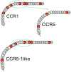
Receptor N-termini representing CCR1, CCR5, and the chimeric CCR5-1like. The CCR1 N-terminus contains a lot of acidic amino acids (outlined in red) in the form of aspartic acid (D) and glutamic acid (E), whereas CCR5 only contains a few. The chimeric receptor CCR5-1like that contains part of the CCR1 receptor N-terminus (ETTTDYDE) resembles CCR1 with regard to acidic amino acid content and spacing, but they are not the same.
Figure 8
Figure 8
Chemokine signaling via CCR5-1like is independent of receptor O-glycosylation status. Ligand-induced Gαi signaling via M1-CCR5 and M1-CCR5-1like in the absence or presence of C21TP81-111 in HEK293 WT, DeltaSia, and sC cells transfected as in Figure 1. (A) As was observed with the untagged CCR5 receptor, compared to CCL5 signaling in WT cells, both potency and efficacy of CCL5 significantly drop in DeltaSia cells and even more so in sC, while C21TP81-111 boosting seems to persist in all cell lines. CCL3- and CCL8-induced signaling via M1-CCR5 is drastically reduced in DeltaSia and sC cells and boosting of CCL3 and CCL8 is absent. (B) In contrast, CCL5, CCL3, and CCL8 signaling via M1-CCR5-1like persists in DeltaSia and sC to almost the same high level as observed in WT cells. C21TP81-111 boosting of CCL5 signaling persists in all three cell lines but is absent for CCL3 and CCL8. Statistical significances were determined by two-way ANOVA with Sidak’s multiple comparisons test (n = 3). ** p ≤ 0.01, * p ≤ 0.05, ns = non-significant.
Figure 9
Figure 9
CCR5-1like retains specificity towards CCR5 cognate ligands. Ligand-induced Gαi signaling via M1-CCR5, M1-CCR5-1like, and M1-CCR1 in HEK293 WT cells transfected as in Figure 1. As can be seen from the left and middle panels, CCR5 and CCR5-1like maintain the same signaling pattern, responding to CCR5 cognate ligands CCL3, CCL5, and CCL8, but not to CCL2, CCL7, and CCL19, as expected. In contrast CCR1, as expected, responds to a broader range of chemokines, including its cognate ligands CCL3, CCL5, and CCL7, but also CCL8 and to a minor degree CCL2. Statistical significances were determined by two-way ANOVA with Sidak’s multiple comparisons test (n = 3). ** p ≤ 0.01, * p ≤ 0.05, ns = non-significant.
Figure 10
Figure 10
Signaling by CCL5 obligate dimer/tetramer versions is boosted similar to WT CCL5 ligand. Ligand-induced Gαi signaling via CCR5 in HEK293 WT cells transfected as in Figure 1. As can be seen, signaling induced by all three ligands (CCL5, E26A, and E66A) via CCR5 is drastically reduced from WT to DeltaSia to sC cells (A,C,E), whereas all three ligands induce potent signaling responses via CCR1 in all three cell lines (B,D,F). Ligand-induced signaling via both CCR5 and CCR1 is boosted by C21TP81-111 in WT cells, a tendency that remains across in DeltaSia and sC cell lines. Interestingly, E66A signaling via CCR5 is significantly boosted by C21TP81-111 in sC cells. Statistical significances were determined by two-way ANOVA with Sidak’s multiple comparisons test (n = 3). ** p ≤ 0.01, * p ≤ 0.05, ns = non-significant.
Figure 11
Figure 11
Schematic representation of the two-step, two-site model of chemokine–receptor interaction with GAG and basic peptide modulation. Due to their negatively charged nature, GAGs on the cell surface and in ECM sequester basic chemokines, creating localized chemokine stores (arrow 1) and less chemokine is captured by the CRS1 (stippled line 1 crossed out). In contrast to chemokine receptors with acidic N-termini, e.g., CCR1, receptors with less acidic N-termini, such as CCR5, rely on post-translational modifications (PTMs) to increase their net negative charge for effective ligand capture (CS1:CRS1 interaction). In the absence of GAGs, native CCR5 signals potently, probably because the surroundings are less acidic, allowing for CCR5 to compete better for ligand capture (arrow 2), as chemokines do not bind strongly to GAG-free cell surface (stippled line 2 crossed out). As published previously, basic peptides, like the C-terminal peptide of CCL21 (C21TP), binds in a dose-dependent manner to acidic receptor N-termini (e.g., CCR7). In the current study we have shown that basic peptides, including C21TP, bind to a peptide corresponding to the unmodified native CCR1 N-terminus but not a peptide corresponding to the unmodified native CCR5 N-terminus. Basic peptides seem to aid the transition from CS1 interaction to CS2 insertion by shielding the acidic receptor N-terminus, allowing for an easier transition from CS1:CRS1 to CS2: CRS2 interaction mode.

Similar articles

References

    1. Bachelerie F., Ben-Baruch A., Burkhardt A.M., Combadiere C., Farber J.M., Graham G.J., Horuk R., Sparre-Ulrich A.H., Locati M., Luster A.D., et al. International Union of Basic and Clinical Pharmacology. [corrected]. LXXXIX. Update on the extended family of chemokine receptors and introducing a new nomenclature for atypical chemokine receptors. Pharmacol. Rev. 2013;66:1–79. doi: 10.1124/pr.113.007724. - DOI - PMC - PubMed
    1. Steen A., Larsen O., Thiele S., Rosenkilde M.M. Biased and g protein-independent signaling of chemokine receptors. Front. Immunol. 2014;5:277. doi: 10.3389/fimmu.2014.00277. - DOI - PMC - PubMed
    1. Corbisier J., Gales C., Huszagh A., Parmentier M., Springael J.Y. Biased signaling at chemokine receptors. J. Biol. Chem. 2015;290:9542–9554. doi: 10.1074/jbc.M114.596098. - DOI - PMC - PubMed
    1. Metzemaekers M., Van D.J., Mortier A., Proost P. Regulation of Chemokine Activity—A Focus on the Role of Dipeptidyl Peptidase IV/CD26. Front. Immunol. 2016;7:483. doi: 10.3389/fimmu.2016.00483. - DOI - PMC - PubMed
    1. Leach K., Charlton S.J., Strange P.G. Analysis of second messenger pathways stimulated by different chemokines acting at the chemokine receptor CCR5. Biochem. Pharmacol. 2007;74:881–890. doi: 10.1016/j.bcp.2007.06.019. - DOI - PubMed

LinkOut - more resources