Review
. 2024 Oct 6;24(19):6458.
doi: 10.3390/s24196458.

Overview of the Design and Application of Photothermal Immunoassays

Affiliations
Review

Overview of the Design and Application of Photothermal Immunoassays

Fengli Gao et al. Sensors (Basel). .

Abstract

Developing powerful immunoassays for sensitive and real-time detection of targets has always been a challenging task. Due to their advantages of direct readout, controllable sensing, and low background interference, photothermal immunoassays have become a type of new technology that can be used for various applications such as disease diagnosis, environmental monitoring, and food safety. By modification with antibodies, photothermal materials can induce temperature changes by converting light energy into heat, thereby reporting specific target recognition events. This article reviews the design and application of photothermal immunoassays based on different photothermal materials, including noble metal nanomaterials, carbon-based nanomaterials, two-dimensional nanomaterials, metal oxide and sulfide nanomaterials, Prussian blue nanoparticles, small organic molecules, polymers, etc. It pays special attention to the role of photothermal materials and the working principle of various immunoassays. Additionally, the challenges and prospects for future development of photothermal immunoassays are briefly discussed.

Keywords: immunoassays; nanozymes; noble metal nanomaterials; photothermal.


Conflict of interest statement

The authors declare no conflicts of interest.

Figures

Figure 3
Figure 3
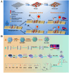
(A) Schematic illustration of an Au@Pt nanostars-based dual-signal LFIA for colorimetric and photothermal detection of SARS-CoV-2 nucleocapsid antibody [71]. Copyright 2024 Elsevier. (B) Schematic illustration of a multimodal LFIA for DHEA detection using multifunctional Au@Pt@Ag NPs with color-photothermal-Raman properties [73]. Copyright 2022 Elsevier. (C) Schematic illustration of a colorimetric and photothermal LFIA based on spark-type AuCuPt alloy for the detection of estriol [75]. Copyright 2024 American Chemical Society. (D) Schematic illustration of (a) photothermal performance comparison of bimetallic nanocuboid Pt3Sn and Pt NPs; (b) detection of S. typhimurium in dairy product samples; (c) principle for the detection of S. typhimurium Using Pt3Sn-based LFIA [77]. Copyright 2024 American Chemical Society.
Figure 8
Figure 8
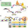
(A) Schematic illustration of a MGO@Au-NCs-based photothermal and colorimetric immunochromatographic biosensor for dual-signal detection of ENR in food samples [109]. Copyright 2024 American Chemical Society. (B) Schematic illustration of (A) preparation of Ab2-modified AgCo@NC NP; (B) construction of a photothermal immunization platform; (C) construction of electrochemical immunization platform [113]. Copyright 2023 American Chemical Society.
Figure 9
Figure 9
(A) Schematic illustration of a black phosphorus-based colorimetric and photothermal immunochromatography for dual-signal detection of NFX [117]. Copyright 2021 Elsevier. (B) Schematic illustration of a photothermal LFIA for the detection of veterinary antibiotic enrofloxacin (ENR) based on AuNPs-enhanced BPNSs [122]. Copyright 2019 Elsevier. (C) Schematic illustration of a dual-signal immunoassay for diethylstilbestrol detection based on BP-Au nanohybrids and the system of TMB-H2O2 [123]. Copyright 2021 Elsevier. (D) Schematic illustration of an immunochromatographic assay for diethylstilbestrol detection using violet phosphorus nanosheets as photothermal materials [125]. Copyright 2023 Elsevier.
Figure 12
Figure 12
(A) Schematic illustration of a photoelectrochemical and photothermal immunoassay for CEA detection based on Ag/MoO3–Pd-mediated gasochromic reactions [153]. Copyright 2023 Elsevier. (B) Schematic illustration of the colorimetric and photothermal dual-signal immunoassay based on MnO2-Au for AOZ detection [154]. Copyright 2022 Elsevier. (C) Schematic illustration of a photothermal immunoassay for the detection of S. typhimurium based on the photothermal effect of magnetic NPs [157]. Copyright 2022 Elsevier.
Figure 16
Figure 16
(A) Schematic illustration of a S. typhimurium strip based on the photothermal effect and catalytic color overlap of PB@Au nanocomposite [119]. Copyright 2022 Elsevier. (B) Schematic illustration of a photothermal immunoassay for pancreatic cancer biomarker CA 19-9 using PBNPs-encapsulated CaCO3 microspheres as signal labels [184]. Copyright 2020 Elsevier. (C) Schematic illustration of a colorimetric and photothermal LFIA for PSA detection based on forcibly dispersed PBNPs [185]. Copyright 2024 American Chemical Society. (D) Schematic illustration of an in situ formed PBNPs-mediated multi-signal nanozyme-linked immunosorbent assay for AFB1 detection [191]. Copyright 2021 American Chemical Society.
Figure 21
Figure 21
(A) Schematic illustration of the preparation of the PCu@pAb probe and sandwich mechanism for the peroxidase-like colorimetric and photothermal multimodal analysis of A. flavus [232]. Copyright 2019 American Chemical Society. (B) Schematic illustration of the photothermal immunosensor using the Cu3(PO4)2/PDA nanosheets [234]. Copyright 2019 American Chemical Society. (C) Schematic illustration of an ICP-MS and photothermal immunoassay for the detection of exosomes based on the bioetching of MnO2 shell and in situ generation of PDA [236]. Copyright 2021 American Chemical Society. (D) Schematic illustration of a TPP-Pdots-based photoelectrochemical and photothermal immunoassay for sensitive detection of sialic acid [237]. Copyright 2019 Elsevier.
Figure 1
Figure 1
(A) Schematic illustration of a rapid and quantitative LFIA using AuNPs as photothermal agents [51]. Copyright 2019 Elsevier. (B) Schematic illustration of a smartphone-integrated photothermal LFIA using dual AuNPs conjugates as labels [53]. Copyright 2024 American Chemical Society.
Figure 2
Figure 2
(A) Schematic illustration of this novel MCI-LFIA biosensor based on AuNF-PMBA NMs for diagnosis of bacterial UTI. (a): Preparation of AuNF–PMBA NMs. (b): Principle of MCI-LFIA for bacterial detection [63]. Copyright 2023 Elsevier. (B) Schematic illustration of a photothermal and colorimetric LFIA for dual-mode determination of SARS-CoV-2 N protein using Au nanoshell-coated Fe3O4 nanoclusters [67]. Copyright 2023 Elsevier.
Figure 4
Figure 4
Schematic illustration of a photothermal immunoassay for CEA detection based on Cu2+-catalyzed consumption of Cys and Cys-induced aggregation of AuNPs [85]. Copyright 2021 Elsevier.
Figure 5
Figure 5
(A) Schematic illustration of a photothermal immunoassay for OTA detection based on the in situ growth of AuNBPs [91]. Copyright 2023 American Chemical Society. (B) Schematic illustration of a colorimetric and photothermal immunoassay for PSA detection through enzyme-triggered growth of AuNS [94]. Copyright 2019 American Chemical Society.
Figure 6
Figure 6
(A) Schematic illustration of fabrication of a CNT-based photothermal immunoassay for the detection of LipL32 antigen [96]. Copyright 2024 American Chemical Society. (B) Schematic illustration of thermal-responsive hydrogel-based multi-signal readout sensing interface for ZEN detection [97]. Copyright 2020 Elsevier.
Figure 7
Figure 7
(A) Schematic illustration of a portable immune-thermometer assay for the detection of S. typhimurium based on the photothermal effect of GO [101]. Copyright 2019 Elsevier. (B) Schematic illustration of an immune-GO-magnetic microbead complex for the development of photothermal immunoassay of cancer cell detection [102]. Copyright 2016 American Chemical Society.
Figure 10
Figure 10
(A) Schematic illustration of a smartphone-based photothermal LFIA for the detection of human anti-SARS-CoV-2 S protein IgG antibodies using ReSe2 nanosheets [133]. Copyright 2023 American Chemical Society. (B) Schematic illustration of an immunochromatography for colorimetric and photothermal detection of nitrofurazone metabolites using the assembly of MoS2@AuNRs as the dual-signal probes [138]. Copyright 2023 Elsevier.
Figure 11
Figure 11
Schematic illustration of an electrochemiluminescence and photothermal immunoassay for the detection of LSR using MXene-based nanocomposite as dual-functional labels [145]. Copyright 2024 Elsevier.
Figure 13
Figure 13
(A) Schematic illustration of Cu2-xSe-Au nanohybrid-based photothermal LFIA for simultaneous detection of three mycotoxins [166]. Copyright 2023 Elsevier. (B) Schematic illustration of dual-plasmonic CuS@Au heterojunctions-based photothermal and colorimetric LFIA for multiplexed detection of T-2 toxin and DON [167]. Copyright 2024 Elsevier.
Figure 14
Figure 14
(A) Schematic illustration of a photothermal and polarity-switchable photoelectrochemical immunoassay for dual-signal detection of CEA based on cation exchange reaction-mediated in situ generation of CuS [171]. Copyright 2023 American Chemical Society. (B) Schematic illustration of a multi-mode immunoassay for PSA detection based on the enzymatic catalysis-induced MOF-confined plasmonic nanozyme [172]. Copyright 2024 American Chemical Society.
Figure 15
Figure 15
(A) Schematic illustration of a photothermal immunoassay of AFB using plasmonic Cu2-xSe NPs-loaded liposomes as photothermal soft nanoballs [173]. Copyright 2019 American Chemical Society. (B) Schematic illustration of a photothermal immunoassay for CEA detection used based on the in situ transformation of Cu2O into Cu31S16 with photothermal effect [176]. Copyright 2022 American Chemical Society.
Figure 17
Figure 17
(A) Schematic illustration of the photothermal-thermoelectric coupled immunoassay for AFP detection with a self-powered temperature sensor [199]. Copyright 2020 American Chemical Society. (B) Schematic illustration of a 1064 nm-excited photothermal immunoassay for PSA detection based on NIR-II-absorbing TMB derivative as the HRP substrate [201]. Copyright 2024 American Chemical Society.
Figure 18
Figure 18
(A) Schematic illustration of the photothermal and colorimetric immunoassay based on the photothermal effect of the iron oxide NPs-mediated TMB-H2O2 colorimetric system [206]. Copyright 2018 American Chemical Society. (B) Schematic illustration of a multi-signal LFIA for the detection of S. aureus using bimetallic Pd/Pt NPs [220]. Copyright 2024 Elsevier.
Figure 19
Figure 19
(A) Schematic illustration of the constructed portable photothermal-colorimetric dual-modality biosensor for cTnI detection [227]. Copyright 2022 Elsevier. (B) Schematic illustration of CoFe PBAs/WS2 nanozyme-based chemiluminescence and photothermal dual-signal LFIA for sensitive detection of gentamicin [228]. Copyright 2024 Elsevier.
Figure 20
Figure 20
(A) Schematic illustration of a split-type proximity hybridization-based photothermal and electrochemiluminescence immunoassay for HE4 detection based on MoS2 NSs and ABTS [229]. Copyright 2020 Elsevier. (B). Schematic illustration of a portable multi-signal immunoassay for the detection of human anti-ASGPR based on plasmonic MXene-induced signal amplification [230]. Copyright 2022 Elsevier.
Figure 22
Figure 22
Schematic illustration of a “three-in-one” multifunctional vanadium-based hollow nanocages with colorimetric, photothermal, and catalytic activities for multi-signal immunoassay [244]. Copyright 2024 American Chemical Society.

Similar articles

References

    1. Jones A., Dhanapala L., Kankanamage R.N.T., Kumar C.V., Rusling J.F. Multiplexed immunosensors and immunoarrays. Anal. Chem. 2020;92:345–362. doi: 10.1021/acs.analchem.9b05080. - DOI - PMC - PubMed
    1. Turner A.P. Biosensors: Sense and sensibility. Chem. Soc. Rev. 2013;42:3184–3196. doi: 10.1039/c3cs35528d. - DOI - PubMed
    1. Pei X., Zhang B., Tang J., Liu B., Lai W., Tang D. Sandwich-type immunosensors and immunoassays exploiting nanostructure labels: A review. Anal. Chim. Acta. 2013;758:1–18. doi: 10.1016/j.aca.2012.10.060. - DOI - PubMed
    1. Pan R., Li G., Liu S., Zhang X., Liu J., Su Z., Wu Y. Emerging nanolabels-based immunoassays: Principle and applications in food safety. TrAC-Trend. Anal. Chem. 2021;145:116462–116482. doi: 10.1016/j.trac.2021.116462. - DOI
    1. Majdinasab M., Lamy de la Chapelle M., Marty J.L. Recent progresses in optical biosensors for interleukin 6 detection. Biosensors. 2023;13:898. doi: 10.3390/bios13090898. - DOI - PMC - PubMed

LinkOut - more resources