. 2024 Sep 26;13(19):2701.
doi: 10.3390/plants13192701.

Establishment of a Sensitive and Reliable Droplet Digital PCR Assay for the Detection of Bursaphelenchus xylophilus

Affiliations

Establishment of a Sensitive and Reliable Droplet Digital PCR Assay for the Detection of Bursaphelenchus xylophilus

Yu Su et al. Plants (Basel). .

Abstract

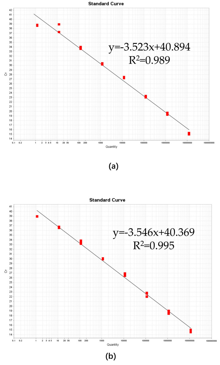
Pine wilt disease (PWD), which poses a significant risk to pine plantations across the globe, is caused by the pathogenic agent Bursaphelenchus xylophilus, also referred to as the pine wood nematode (PWN). A droplet digital PCR (ddPCR) assay was developed for the quick identification of the PWN in order to improve detection sensitivity. The research findings indicate that the ddPCR assay demonstrated significantly higher analysis sensitivity and detection sensitivity in comparison to traditional quantitative PCR (qPCR). However, it had a more limited dynamic range. High specificity was shown by both the ddPCR and qPCR techniques in the diagnosis of the PWN. Assessments of reproducibility revealed that ddPCR had lower coefficients of variation at every template concentration. Inhibition tests showed that ddPCR was less susceptible to inhibitors. There was a strong linear association between standard template measurements obtained using ddPCR and qPCR (Pearson correlation = 0.9317; p < 0.001). Likewise, there was strong agreement (Pearson correlation = 0.9348; p < 0.001) between ddPCR and qPCR measurements in the evaluation of pine wood samples. Additionally, wood samples from symptomatic (100% versus 86.67%) and asymptomatic (31.43% versus 2.9%) pine trees were diagnosed with greater detection rates using ddPCR. This study's conclusions highlight the advantages of the ddPCR assay over qPCR for the quantitative detection of the PWN. This method has a lot of potential for ecological research on PWD and use in quarantines.

Keywords: Bursaphelenchus xylophilus determination; droplet digital PCR; nuclear DNA.


Conflict of interest statement

The authors declare no conflict of interest.

Figures

Figure 1
Figure 1
Alignment of the consensus 16SrDNA sequence from the B. xylophilus (Bx) and B. mucronstus (Bm) isolates. The forward primer and reverse primer are indicated by red arrows, and the probe is indicated by a red box. The consensus DNA sequence from B. xylupholus and B. mucronstus is highlighted with a dark-blue background. Dots indicate gaps in the corresponding sequence that were added for the alignment.
Figure 2
Figure 2
Optimization of the qPCR and ddPCR assays. (a) The amplification plot of the qPCR assay displays the optimal extension temperature for the detection of B. xylophilus. The unbroken blue line represents the threshold automatically set by the software. The reactions occurred across the following extension temperature gradient: 50 °C, 53 °C, 56 °C, 59 °C, 62 °C, and 65 °C. Different extension temperatures exhibited slight variations in Ct values. (b) Fluorescence amplitudes are plotted against an annealing temperature gradient. Positive droplets (blue) with PCR amplification are shown above the unbroken blue line, and negative droplets (black) without any amplification are shown below the line. Six ddPCR reactions, each containing the same number of targets, are separated by a vertical gray dotted line. The reactions occurred across the following annealing temperature gradient: 48 °C, 51 °C, 54 °C, 57 °C, 60 °C, and 63 °C. Different annealing temperatures exhibited slight distinctions in fluorescence between negative and positive droplets, and the highest fluorescence intensity was observed at 57 °C.
Figure 3
Figure 3
Specificity of qPCR (a) and ddPCR (b) assays for B. xylophilus. Amplification was conducted with DNA templates from B. xylophilus (BX01-BX06), B. mucronatus (BM01-BM03), B. thailandae (BT01-BT03), B. sexdentati (BS01), B. dalianensis (BD01), Aphelenchoides parasaprophilus (AS01), A. resinosi (AR01 and AR02), and Cylindrotylenchus pini (CP01).
Figure 4
Figure 4
The standard curves of the qPCR assays, which were generated using plasmid DNA (a) and samples spiked with pine wood DNA extract (b). Plasmid DNA was diluted 10-fold to create eight concentration gradients. The resulting standard curve for the plasmid DNA had a slope of −3.523, corresponding to a PCR efficiency of 92.246% (R2 = 0.989). For the spiked samples, the standard curve exhibited a slope of −3.546, which was equivalent to a PCR efficiency of 91.419% (R2 = 0.995).
Figure 5
Figure 5
Linear regression of the qPCR assay for plasmid DNA (a) and spiked samples (b) using the serial dilution series. Both of these two assays exhibited excellent linearity (both R2 = 0.999).
Figure 6
Figure 6
Linear regression of the ddPCR assay for plasmid DNA (a) and spiked samples (b) constructed using the same serial dilution series tested with the qPCR assay (see Figure 5). The estimated Pearson correlation coefficient of the plasmid DNA regression curve (y = 0.8843x + 0.1733) is 0.9947 (R2 = 0.9991, p < 0.0001). The estimated Pearson correlation coefficient of the regression curve of the spiked samples (y = 0.7876x + 0.5153) is 0.9773 (R2 = 0.9773, p < 0.0001). The standards tested using ddPCR exhibited a dynamic range of five orders of magnitude. The vertical axis shows the log5-transformed copy number/μL of the ddPCR reaction mixture. The horizontal ordinate indicates the expected log5-transformed concentration of the copy number/μL of the ddPCR reaction mixture.
Figure 7
Figure 7
A representative 1-D plot of ddPCR reactions. The ordinate indicates the fluorescence amplitude, and the blue line is the threshold. Above the threshold are positive droplets (blue) containing at least one copy of the target DNA, and below the threshold are negative droplets (gray) without the target DNA. Eight ddPCR reactions with various serially diluted targets are divided by the vertical dotted line. When the target DNA exceeded 106, the ddPCR reaction was saturated by an excess target DNA concentration, and when the target DNA was 10−1, the ddPCR reaction provided a negative result.
Figure 8
Figure 8
The inter-assay CV% of the qPCR and ddPCR assays. Samples P6-1, P6-2, P7-1, P7-2, P8-1, P8-2, P9-1, and P9-2 were positive plasmid DNA; among them, P6-1, P6-2, P7-1, and P7-2 had high concentrations, while P8-1, P8-2, P9-1, and P9-2 had low concentrations. Histograms indicate the average copy number of each sample on a log10 scale. Lines show the CV variation trends of the qPCR and ddPCR assays during repeated tests with diverse sample concentrations. The ddPCR assay was more precise than the qPCR assay for quantification of the PWN, especially for low target concentrations (numerical data supporting Figure 6 are provided in Table S4).
Figure 9
Figure 9
The influence of samples spiked with different volumes of pine wood extracts. ddPCR exhibited superior tolerance to pine wood extracts compared to the qPCR assay. qPCR was more susceptible to the inhibitors (a). In the ddPCR assays, the fluorescence intensity of positive droplets decreased slightly with an increase in the inhibitor volume (b). In the qPCR assays, the fluorescence curves exhibited no significant differences between different volumes of inhibitors (c) (numerical data supporting Figure 9 are provided in Table S5).
Figure 10
Figure 10
Correction of qPCR and ddPCR measurements. Measurements of pine wood samples using qPCR and ddPCR assays were significantly correlated (Pearson r = 0.9348, p < 0.0001). Solid line indicates fitting curve (numerical data from ddPCR and qPCR assays are provided in Table S8).

Similar articles

Cited by

References

    1. Mamiya Y. Pathology of the Pine Wilt Disease Caused by Bursaphelenchus xylophilus. Annu. Rev. Phytopathol. 1983;21:201–220. doi: 10.1146/annurev.py.21.090183.001221. - DOI - PubMed
    1. Nascimento F., Hasegawa K., Mota M., Vicente C. Bacterial Role in Pine Wilt Disease Development—Review and Future Perspectives: Bacteria in Pine Wilt Disease. Environ. Microbiol. Rep. 2014;7:51–63. doi: 10.1111/1758-2229.12202. - DOI - PubMed
    1. Li Y.-L., Fan C.-J., Jiang X.-H., Tian X.-Y., Han Z.-M. Bursaphelenchus xylophilus: An Important Pathogenic Factor of Pine Wilt Disease and Its Relationship with Bursaphelenchus mucronatus. Plant Dis. 2021;105:3055–3062. doi: 10.1094/PDIS-02-21-0396-RE. - DOI - PubMed
    1. Myers R.F. Pathogenesis in Pine Wilt Caused by Pinewood Nematode, Bursaphelenchus xylophilus. J. Nematol. 1988;20:236–244. - PMC - PubMed
    1. Bolla R.I., Weaver C., Winter R.E.K. Genomic Differences among Pathotypes of Bursaphelenchus xylophilus. J. Nematol. 1988;20:309–316. - PMC - PubMed

LinkOut - more resources