. 2024 Oct 8;14(19):2891.
doi: 10.3390/ani14192891.

Morphology of Larger Salivary Glands in Peccaries (Pecari tajacu Linnaeus, 1758)

Affiliations

Morphology of Larger Salivary Glands in Peccaries (Pecari tajacu Linnaeus, 1758)

Carlos Magno Oliveira Júnior et al. Animals (Basel). .

Abstract

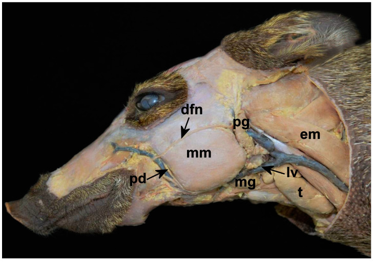
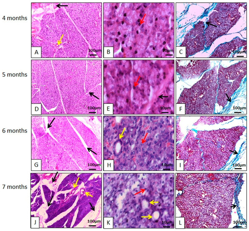
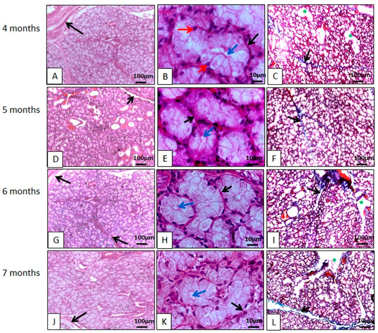
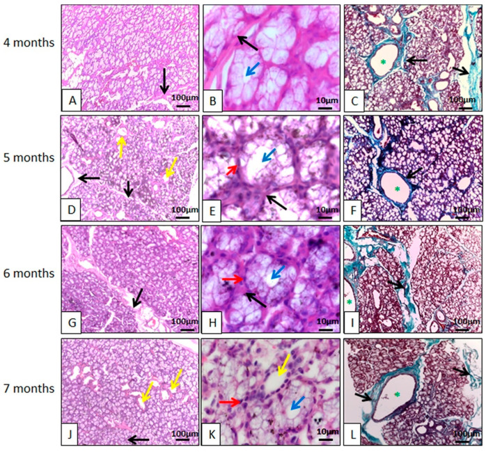
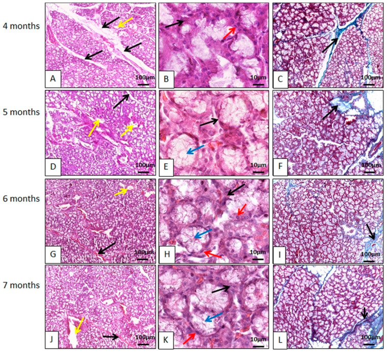
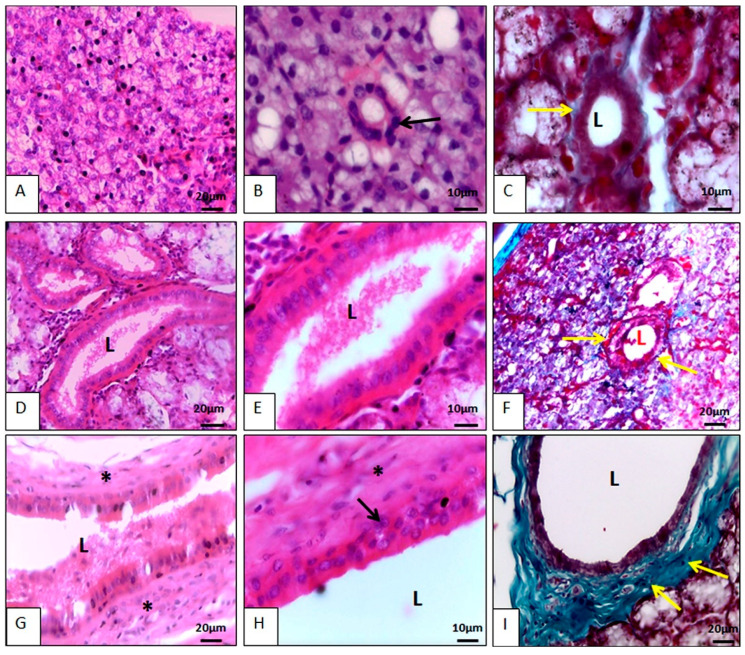
This work aims to study the major salivary gland morphology of peccaries during their growth. The glands were analyzed using macroscopic description, light microscopy, electron microscopy, histochemistry, and immunohistochemistry. Topographically, the salivary glands resemble other animals, including domestic animals and pigs. During growth, the parotid enlarges and mandibular gland loses weight. Histologically, the parotid has serous production, and sublingual has mucous production, resembles most species, however, mandibular gland produces mucous, unlike other animals, including pigs, which produce seromucous secretion. Histochemically, parotid produces more acidic mucins than pigs and it undergoes maturation during development; mandibular, and especially the sublingual gland, produce more acidic and basic mucopolysaccharides than pigs. The results found with transmission and scanning electron microscopy techniques corroborate the histological and histochemistry findings. The major salivary glands were positive to different lecithins (Com-A, BSA-I-B4, WGA and PNA), which were also more positive than in pigs and sheep. We conclude that collared peccaries have a salivary secretion that facilitates the digestion of carbohydrates, and biometric characteristics and positivity to lecithins that facilitate adaptation to foods with antinutritional factors.

Keywords: Tayassuidae; lecithins; ultrastructure; wild animals.


Conflict of interest statement

The authors declare no conflicts of interest.

Figures

Figure 1
Figure 1
Photographic image of the macroscopy of the major salivary glands of the collared peccary (Pecari tajacu Linnaeus, 1758). Mandibular gland (mg) is located ventrally to the linguofacial vein (lv). Above is part of the parotid gland (pg), which is rostrally to the sternocephalicus muscle (em). The “em” is positioned dorsally to the thymus (t). Rostrally we still find the masseter muscle (mm), surrounded dorsally by the dorsal facial nerve (dfn), and caudally by the parotid duct (pd).
Figure 2
Figure 2
Photomicrographs of the parotid gland of the collared peccary (Pecari tajacu Linnaeus, 1758). In (A,D,G,J), it can be seen that the basophilic lobes are individualized by connective tissue (black arrows). In (B,E,H,K), the connective tissue (black arrows) delimits the acini and ducts (yellow arrows). The nuclei are spherical and are strongly stained by hematoxylin (red arrows). In (C,F,I,L), it can be seen that the lobes are individualized by collagen fibers (black arrows). (A,B,D,E,G,H,J,K) Hematoxylin-Eosin staining; (C,F,I,L) Gomori Trichrome Staining.
Figure 3
Figure 3
Photomicrographs of the collared peccary mandibular gland (Pecari tajacu Linnaeus, 1758). In (A,D,G,J), it can be seen that the acidophilic lobes are individualized by connective tissue (black arrows). In (B,E,H,K), connective tissue (black arrows) delimits the acini. The acinar cells are organized to form the acinar lumen (blue arrows). The nuclei of these cells are spherical and stain strongly with hematoxylin (red arrows). In (C,F,I,L), the ducts (*) are individualized by collagen fibers (black arrows). (A,B,D,E,G,H,J,K) Hematoxylin-Eosin staining; (C,F,I,L) Gomori Trichrome staining.
Figure 4
Figure 4
Photomicrographs of the polystomatic sublingual gland of the collared peccary (Pecari tajacu Linnaeus, 1758). In (A,D,G,J), the acini and ducts (yellow arrows) are individualized by connective tissue (black arrows). In (B,E,H,K), the acini and ducts (yellow arrows) are individualized by connective tissue (black arrows). The acinar cells are organized to form the acinar lumen (blue arrows), the nuclei of these cells are spherical and stain strongly with hematoxylin (red arrows). In (C,F,I,L), the connective tissue (black arrows) delimited the acini and ducts (*). (A,B,D,E,G,H,J,K) Hematoxylin-Eosin staining; (C,F,I,L) Gomori Trichrome staining.
Figure 5
Figure 5
Photomicrographs of the monostomatic sublingual gland of the collared peccary (Pecari tajacu Linnaeus, 1758). In (A,D,G,J), the acini and ducts (yellow arrows) are individualized by connective tissue (black arrows). In (B,E,H,K) connective tissue (black arrows) delimit the acini and ducts (yellow arrows), the nuclei are flat, stain strongly with hematoxylin, and are in the basal region (red arrows). The acinar cells are with its apex facing the acinar lumen (blue arrows). In (C,F,I,L), the acini and ducts are individualized by collagen fiber meshes. (A,B,D,E,G,H,J,K) Hematoxylin-Eosin staining; (C,F,I,L) Gomori Trichrome staining.
Figure 6
Figure 6
Photomicrographs of the salivary ducts of the collared peccary (Pecari tajacu Linnaeus, 1758). In (A), the arrangement of the intercalated ducts is observed. There is an intimate relationship between this duct and the acini. In (B), it can be seen that the duct is made up of a single layer of cubic epithelial cells (black arrows) and its nucleus is spherical, basophilic, and located in the cell center. In (C), the duct is delimited by a thin layer of collagen fiber (yellow arrows). In (D), there is the striated duct that was larger than the intercalated duct. In (E), it is observed that the cells were columnar, and the nuclei were spherical and positioned in the center of the cell. Note that the cell base is more eosinophilic than the cell apex. In (F), a thin layer of collagen fiber (yellow arrows) can be seen delimiting the duct. In (G), the excretory duct can be seen. This duct had a lot of connective tissue around it (*) and was in the interlobular region. In (H), it is noted that the excretory duct contained more than one layer of epithelial cells, the most basal of which was cubic (black arrow) and the outermost columnar. In (I), a dense network of collagen fibers is noted surrounding the excretory duct. L = lumen. (A,B,D,E,G,H) Hematoxylin-Eosin staining. (C,F,I) Gomori Trichrome staining.

Similar articles

References

    1. Pedersen A.M., Bardow A., Jensen S.B., Nauntofte B. Saliva and Gastrointestinal Functions of Taste, Mastication, Swallowing, and Digestion. Oral Dis. 2002;8:117–129. doi: 10.1034/j.1601-0825.2002.02851.x. - DOI - PubMed
    1. Mavri G., Škrlep M., Rutland C.S., Potočnik B., Batorek-Lukač N., Kubale V. Salivary Gland Adaptation to Dietary Inclusion of Hydrolysable Tannins in Boars. Animals. 2022;12:2171. doi: 10.3390/ani12172171. - DOI - PMC - PubMed
    1. Denny P., Hagen F.K., Hardt M., Liao L., Yan W., Arellanno M., Bassilian S., Bedi G.S., Boontheung P., Cociorva D., et al. The proteomes of human parotid and submandibular/sublingual gland salivas collected as the ductal secretions. J. Proteome Res. 2008;7:1994–2006. doi: 10.1021/pr700764j. - DOI - PMC - PubMed
    1. Oliveira Júnior C.M., Bezerra F.V.F., Câmara F.V., Vale A.M., Oliveira G.B., Silva A.R., Ambrosio C.E., Oliveira M.F. Morfologia das glândulas salivares maiores em cutias (Dasyprocta leporina Linnaeus, 1766) Pesqui. Veterinária Bras. 2016;36:227–236. doi: 10.1590/S0100-736X2016000300013. - DOI
    1. Poddar S., Jacob S. Gross and Microscopic Anatomy of the Major Salivary Glands of the Ferret. Acta Anat. 1977;98:434–443. doi: 10.1159/000144823. - DOI - PubMed

LinkOut - more resources