Pathological Changes and Sodium Rhodizonate Test as Tools for Investigating Gunshot Wounds in Veterinary Forensic Pathology
- PMID: 39409862
- PMCID: PMC11476102
- DOI: 10.3390/ani14192913
Pathological Changes and Sodium Rhodizonate Test as Tools for Investigating Gunshot Wounds in Veterinary Forensic Pathology
Abstract
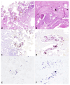
Gunshot wound morphology and gunshot residues (GSRs) evaluation have been poorly investigated in veterinary forensic pathology. The aims of the present study were to assess the gunshot wound morphology in animals and evaluate the detectability of lead deriving from GSRs using colorimetric techniques. To these aims, cadavers were divided into four different groups. Group A comprised eight animals who died from firearm-related injuries, while groups B and C included dog limbs shot using different shooting ranges; group D comprised dog limbs stabbed with a screwdriver. Morphological analysis was performed on all entry gunshot wounds. Lead residues were investigated using a Bullet Hole Testing Kit (BTK) and Rhodizonate Sodium histochemical staining (NaR-s). Gunshot wounds in group A showed an abrasion ring associated with hemorrhages and tissue necrosis. Groups B and C showed injuries related to the shooting range. NaR-s showed positive results in both animals that died from gunshot wounds and experimentally shot limbs. However, the number of positive cases and the pattern of lead distribution varied with the shooting range. Positive results by BTK were limited to close-contact shots in group B limbs. Our results suggest that both pathological examination and NaR tests represent valid tools for investigating gunshot wounds in veterinary pathology.
Keywords: forensic science; gunshot residues; penetrating injuries; veterinary forensic pathology.
Conflict of interest statement
The authors declare no conflicts of interest.
Figures


Similar articles
-
Is it possible to detect lead derived from gunshot residues on decalcified human bone by means of a histochemical staining with sodium rhodizonate?Forensic Sci Int. 2020 Nov;316:110474. doi: 10.1016/j.forsciint.2020.110474. Epub 2020 Sep 1. Forensic Sci Int. 2020. PMID: 32882639
-
Can cadaverous pollution from environmental lead misguide to false positive results in the histochemical determination of Gunshot Residues? In-depth study using ultra-sensitive ICP-MS analysis on cadaveric skin samples.Forensic Sci Int. 2018 Nov;292:23-26. doi: 10.1016/j.forsciint.2018.08.041. Epub 2018 Sep 13. Forensic Sci Int. 2018. PMID: 30268034
-
Detectability and medico-legal value of the gunshot residues in the intracorporeal channel.J Forensic Leg Med. 2015 Nov;36:10-5. doi: 10.1016/j.jflm.2015.08.004. Epub 2015 Aug 21. J Forensic Leg Med. 2015. PMID: 26320004
-
Arrow entrance wounds with blackened margins simulating bullet wipe.Int J Legal Med. 2020 Jan;134(1):283-294. doi: 10.1007/s00414-019-02191-1. Epub 2019 Nov 11. Int J Legal Med. 2020. PMID: 31713063
-
Practical pathology of gunshot wounds.Arch Pathol Lab Med. 2006 Sep;130(9):1283-9. doi: 10.5858/2006-130-1283-PPOGW. Arch Pathol Lab Med. 2006. PMID: 16948512 Review.
References
-
- Lotfollahzadeh S., Burns B. StatPearls. StatPearls Publishing; Treasure Island, FL, USA: 2023. [(accessed on 15 April 2024)]. Penetrating Abdominal Trauma. Available online: https://www.ncbi.nlm.nih.gov/books/NBK459123/
Grants and funding
LinkOut - more resources
Full Text Sources

