

Vascular Biomarkers for Pulmonary Nodule Malignancy: Arteries vs. Veins
- PMID: 39409894
- PMCID: PMC11476001
- DOI: 10.3390/cancers16193274
Vascular Biomarkers for Pulmonary Nodule Malignancy: Arteries vs. Veins
Abstract
Objective: This study aims to investigate the association between the arteries and veins surrounding a pulmonary nodule and its malignancy.
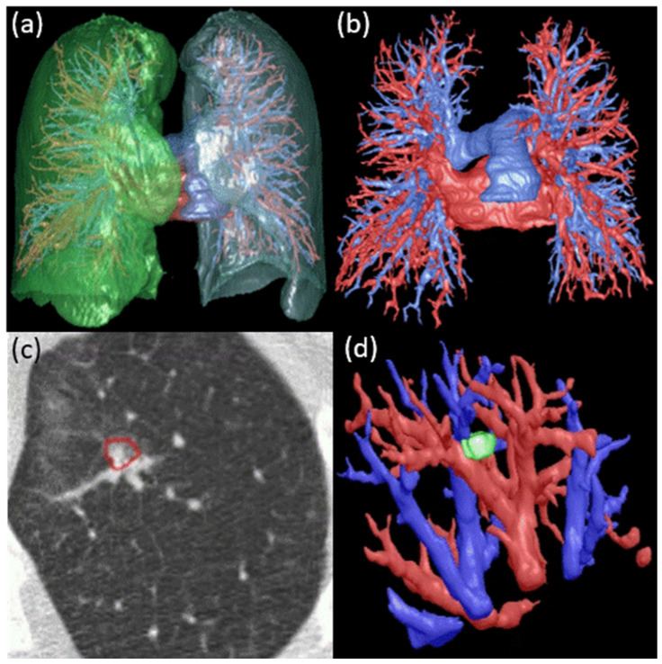
Methods: A dataset of 146 subjects from a LDCT lung cancer screening program was used in this study. AI algorithms were used to automatically segment and quantify nodules and their surrounding macro-vasculature. The macro-vasculature was differentiated into arteries and veins. Vessel branch count, volume, and tortuosity were quantified for arteries and veins at different distances from the nodule surface. Univariate and multivariate logistic regression (LR) analyses were performed, with a special emphasis on the nodules with diameters ranging from 8 to 20 mm. ROC-AUC was used to assess the performance based on the k-fold cross-validation method. Average feature importance was evaluated in several machine learning models.
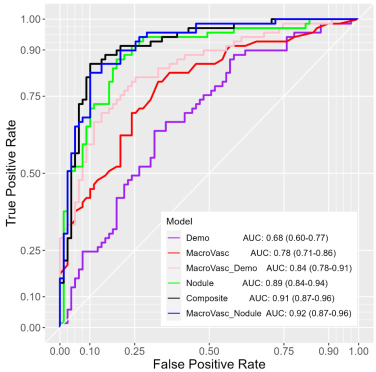
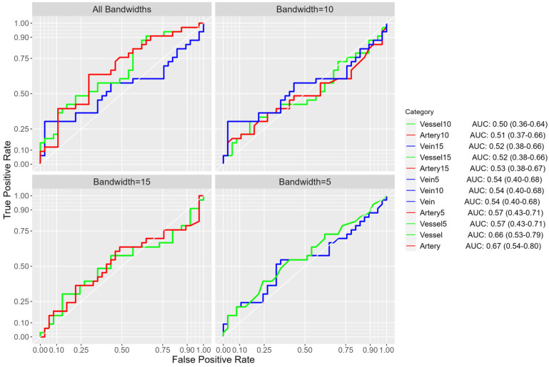
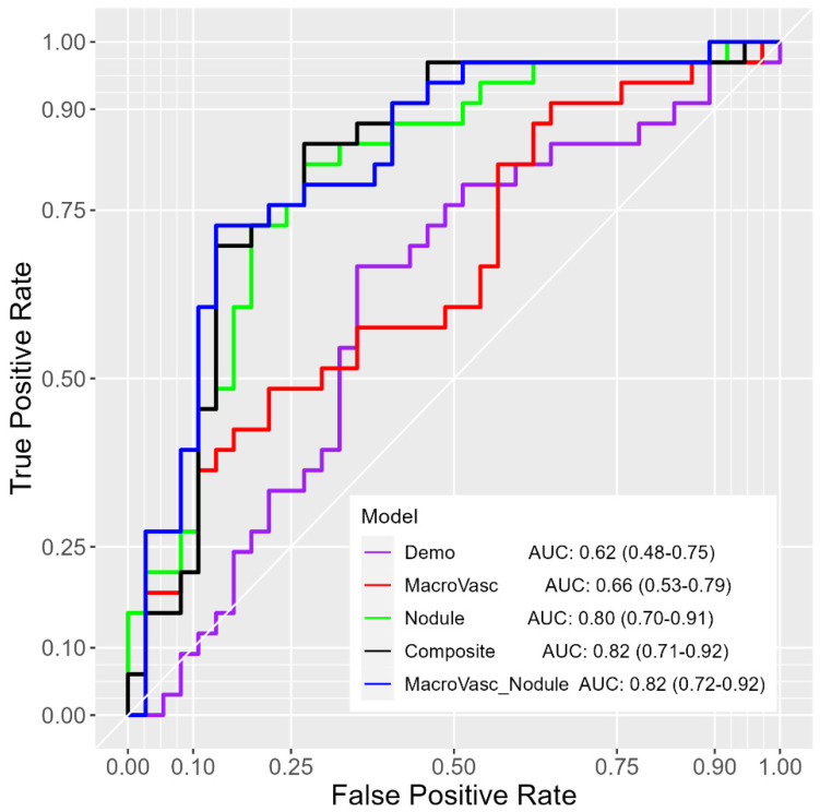
Results: The LR models using macro-vasculature features achieved an AUC of 0.78 (95% CI: 0.71-0.86) for all nodules and an AUC of 0.67 (95% CI: 0.54-0.80) for nodules between 8-20 mm. Models including macro-vasculature features, demographics, and CT-derived nodule features yielded an AUC of 0.91 (95% CI: 0.87-0.96) for all nodules and an AUC of 0.82 (95% CI: 0.71-0.92) for nodules between 8-20 mm. In terms of feature importance, arteries within 5.0 mm from the nodule surface were the highest-ranked among macro-vasculature features and retained their significance even with the inclusion of demographics and CT-derived nodule features.
Conclusions: Arteries within 5.0 mm from the nodule surface emerged as a potential biomarker for effectively discriminating between malignant and benign nodules.
Keywords: machine learning; macro-vasculature; malignancy; pulmonary nodules.
Conflict of interest statement
The authors declare no conflicts of interest.
Figures
References
Grants and funding
LinkOut - more resources
Full Text Sources
