. 2024 Sep 26;16(19):3277.
doi: 10.3390/cancers16193277.

Xeroderma Pigmentosum Type C Primary Skin Fibroblasts Overexpress HGF and Promote Squamous Cell Carcinoma Invasion in the Absence of Genotoxic Stress

Affiliations

Xeroderma Pigmentosum Type C Primary Skin Fibroblasts Overexpress HGF and Promote Squamous Cell Carcinoma Invasion in the Absence of Genotoxic Stress

Sahar Al-Qaraghuli et al. Cancers (Basel). .

Abstract

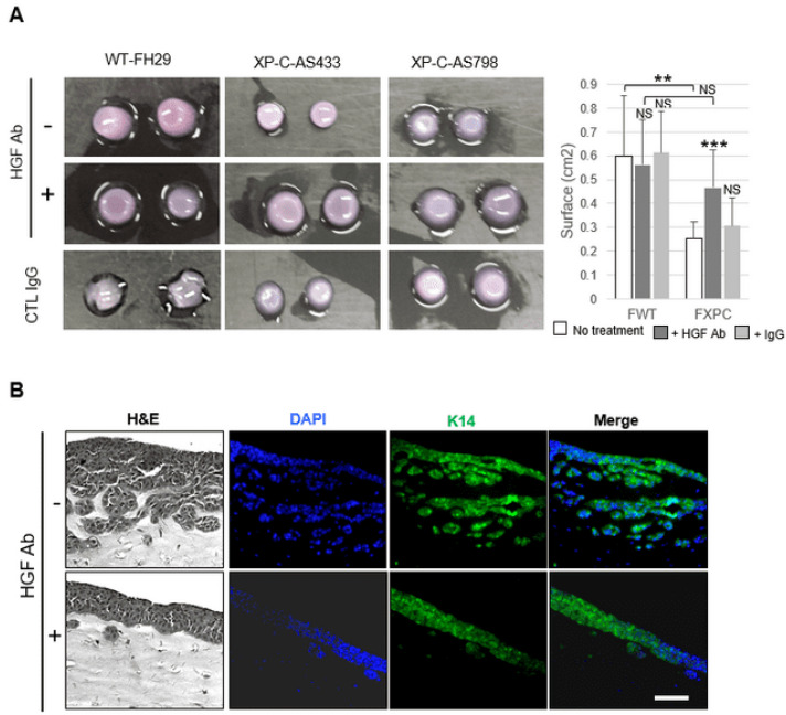
Xeroderma pigmentosum (XP) is a very rare recessive disease caused by the incapacity to resolve ultraviolet-induced DNA lesions through Nucleotide Excision Repair (NER). Most XP patients suffer from aggressive skin carcinoma and melanoma at a very early age (<8). Our previous results showed that primary XP fibroblasts isolated from healthy (non-photo-exposed) skin negatively impact the extracellular matrix and fail to activate the innate immune system. Here, we show for the first time that XP-C fibroblasts also play a major role in cancer cell invasion ex vivo and in vivo through the overexpression of Hepatocyte Growth Factor/Scatter Factor (HGF/SF) in the absence of genotoxic attacks. The use of inhibitors of the activation of the HGF/SF pathway counteracted the effects of XP fibroblasts on the growth of cancer cells, suggesting new perspectives in the care of XP patients.

Keywords: HGF/SF; fibroblasts; squamous cell carcinoma; xeroderma pigmentosum.


Conflict of interest statement

The authors declare no conflict of interest.

Figures

Figure 1
Figure 1
XP-C fibroblasts promote cancer cell invasion in organotypic skin cultures. (A) Representative images of H&E staining of sections from organotypic cultures generated from either WT keratinocytes (KTM1) or SCC13 cells together with either WT (FH29) or XP-C (AS433, AS798) fibroblasts embedded in dermal equivalents; bar: 50 µm. (B) Quantification of SSC13 cell invasion. Values are presented as the mean ± SD of three independent areas; *** p < 0.0001. (C) Immunofluorescence staining of K14 keratin (green) and nuclei (blue) show that invading cells are of epithelial origin; bar: 50 µm. (D) Spheroid assays composed of SCC13-GFP cells and either (red) WT (FH29) or XP-C (AS798) fibroblasts; bar: 250 µm. (E) Confocal microscopy images of spheroids showing SCC13-GFP cells and XP-C fibroblasts. Insets show higher magnification of spheroids. Arrowheads indicate the XP-C fibroblasts leading to invading protrusions. Bar: 40 µm.
Figure 2
Figure 2
XP-C fibroblasts increase the rate of scratch closure of cancer cells ex vivo. Effects of either WT or XP-C fibroblast CM on SCC cells’ scratch closure without (A) or with MMC (B). Upper panels show representative micrografts from the time-lapse video microscopy of SCC13 cells’ scratch closure in the presence of either WT or XP-C CM at indicated times. Lower panels show the kinetics of SCC13 cells’ scratch closure in the presence of either 2 strains of XP-C CM (XP-C-AS433, XP-C-AS798) or 1 strain of WT fibroblast CM (WT-FH29). Dash lines indicate merging of the scratch. Note the substantially slower scratch closure kinetics of SCC13 cells in the presence of MMC (please also see Figure S2).
Figure 3
Figure 3
XP-C fibroblasts overexpress HGF/SF, leading to downstream activation of STAT3 and JNK c-MET pathways in SCC cells. (A) Accumulation of HGF mRNA was significantly higher in XP-C (n = 4) (FXP-C) than in WT (n = 2) fibroblasts (FWT). Values are presented as the mean ± SD of 4 independent experiments, *** p < 0.001. (B) HGF levels (ELISA) were significantly higher in XP-C fibroblast CM (n = 3) than in WT fibroblast CM (n = 3); ** p < 0.01. (C) Immunoblot analysis of cMET and p-MET in SCC cells treated with CM of either WT (FH29, FH84) or XP-C (AS148, AS433, AS875, AS798) fibroblasts. (DG) Western blot analysis of phosphorylated forms of STAT3, JNK1,2, ERK1, 2, and P38 in SCC13 cells cultured in CM of WT (FTM5, FH29) or XP-C (AS148, AS433, AS875, AS798) primary fibroblasts. β-tubulin was used as the loading/transfer control. Right panel quantifies the ratio of phosphorylated to total protein amounts. Values are the mean ± SD of two independent experiments. *** p < 0.001, * p < 0.05.
Figure 4
Figure 4
Blockade of cMET/HGF in culture media conditioned by primary XP-C fibroblasts reduces activation of JUNK and STAT3. (A) Western blot analysis of total cMET and p-MET levels in lysates of SCC cells cultured in XP-C fibroblast CM (AS875, AS798) ± HGF blocking antibody. GAPDH was used as the loading/transfer control. Right panel shows quantification of p-MET accumulation ± anti HGF-blocking antibody. (B) Western blot analysis of p-STAT3 and p-JNK levels in lysates of SCC cells cultured in the presence of XP-C fibroblast CM (AS798) ± HGF blocking antibody. Tubulin was used as the loading/transfer control. Right panel shows the quantification of the ratio of p-STAT3 and p-JNK to the total protein levels under the different conditions. Values are the mean ± SD. *** p < 0.001; * p < 0.05). For Figure 3 and Figure 4, raw images of blots are shown in Figure S4.
Figure 5
Figure 5
Contractile and invasive properties of cancer cells are normalized by HGF antibodies in XP-C CM. (A) Organoids containing either WT (FH29) or XP-C (AS433, AS798) fibroblasts ± HGF blocking antibody; quantitation is showed in the right panel. (B) Organotypic cultures composed of SCC cells and XP-C (AS798) fibroblasts ± HGF blocking antibody. Sections were stained using either H&E, DAPI (blue), or K14 antibody (green). Cancer cell invasion was significantly reduced by the HGF antibody; bar: 50 µm. Values are the mean ± SD. ** p < 0.01, *** p < 0.001.
Figure 6
Figure 6
Contribution of XP-C fibroblasts to carcinoma cell invasion in vivo. (A) Numbers and tumor volume were measured in either absence or presence of XP-C fibroblasts. (B) In contrast to WT primary fibroblasts (FTM1, FTM5, FH84, FH29; representative image in upper panel), XP-C primary fibroblasts (AS 433, AS 875, AS 875, AS 673, AS 202; lower panel) accumulated in the vicinity of epithelial cancer cell invasions (representative image of graft sections from xenografts). Labeling colors are indicated; bar: 100 µm.
Figure 7
Figure 7
XP-C primary fibroblasts accumulate higher ROS levels than WT primary fibroblasts after treatment with either H2O2 or UVA, as indicated in the figure. Pool of 2 independent experiments using WT FH 84 primary fibroblasts as controls; WT, n = 2; XP-C, n = 3. Asterisks indicate statistical significance: ** p < 0.01, *** p < 0.001.

Similar articles

References

    1. Bray F., Ferlay J., Soerjomataram I., Siegel R.L., Torre L.A., Jemal A. Global cancer statistics 2018: GLOBOCAN estimates of incidence and mortality worldwide for 36 cancers in 185 countries. CA Cancer J. Clin. 2018;68:394–424. doi: 10.3322/caac.21492. - DOI - PubMed
    1. Bradford P.T., Goldstein A.M., Tamura D., Khan S.G., Ueda T., Boyle J., Oh K.S., Imoto K., Inui H., Moriwaki S., et al. Cancer and neurologic degeneration in xeroderma pigmentosum: Long term follow-up characterises the role of DNA repair. J. Med. Genet. 2011;48:168–176. doi: 10.1136/jmg.2010.083022. - DOI - PMC - PubMed
    1. Farkas G., Kocsis Z.S., Szekely G., Dobozi M., Kenessey I., Polgar C., Juranyi Z. Smoking, chromosomal aberrations, and cancer incidence in healthy subjects. Mutat. Res. Genet. Toxicol. Environ. Mutagen. 2021;867:503373. doi: 10.1016/j.mrgentox.2021.503373. - DOI - PubMed
    1. Chung J.H., Seo J.Y., Choi H.R., Lee M.K., Youn C.S., Rhie G., Cho K.H., Kim K.H., Park K.C., Eun H.C. Modulation of skin collagen metabolism in aged and photoaged human skin in vivo. J. Investig. Dermatol. 2001;117:1218–1224. doi: 10.1046/j.0022-202x.2001.01544.x. - DOI - PubMed
    1. de Feraudy S., Boubakour-Azzouz I., Fraitag S., Berneburg M., Chan L., Chew K., Clericuzio C.L., Cunningham B., Tope W.D., Cleaver J.E. Diagnosing xeroderma pigmentosum group C by immunohistochemistry. Am. J. Dermatopathol. 2010;32:109–117. doi: 10.1097/DAD.0b013e3181af0a5e. - DOI - PubMed

LinkOut - more resources