. 2024 Sep 26;16(19):3286.
doi: 10.3390/cancers16193286.

Impact of Optimized Ku-DNA Binding Inhibitors on the Cellular and In Vivo DNA Damage Response

Affiliations

Impact of Optimized Ku-DNA Binding Inhibitors on the Cellular and In Vivo DNA Damage Response

Pamela L Mendoza-Munoz et al. Cancers (Basel). .

Abstract

Background: DNA-dependent protein kinase (DNA-PK) is a validated cancer therapeutic target involved in DNA damage response (DDR) and non-homologous end-joining (NHEJ) repair of DNA double-strand breaks (DSBs). Ku serves as a sensor of DSBs by binding to DNA ends and activating DNA-PK. Inhibition of DNA-PK is a common strategy to block DSB repair and improve efficacy of ionizing radiation (IR) therapy and radiomimetic drug therapies. We have previously developed Ku-DNA binding inhibitors (Ku-DBis) that block in vitro and cellular NHEJ activity, abrogate DNA-PK autophosphorylation, and potentiate cellular sensitivity to IR. Results and Conclusions: Here we report the discovery of oxindole Ku-DBis with improved cellular uptake and retained potent Ku-inhibitory activity. Variable monotherapy activity was observed in a panel of non-small cell lung cancer (NSCLC) cell lines, with ATM-null cells being the most sensitive and showing synergy with IR. BRCA1-deficient cells were resistant to single-agent treatment and antagonistic when combined with DSB-generating therapies. In vivo studies in an NSCLC xenograft model demonstrated that the Ku-DBi treatment blocked IR-dependent DNA-PKcs autophosphorylation, modulated DDR, and reduced tumor cell proliferation. This represents the first in vivo demonstration of a Ku-targeted DNA-binding inhibitor impacting IR response and highlights the potential therapeutic utility of Ku-DBis for cancer treatment.

Keywords: DNA-PK; Ku-DBis; double-strand break repair; non-homologous end joining; non-small cell lung cancer; small-molecule inhibitors.


Conflict of interest statement

Author Karim Ben Ali Gacem was employed by the company Sanofi. J.J. Turchi is a co-founder and CSO of NERx Biosciences. J.J. Turchi and N.S. Gavande are co-inventors listed on patents covering the compounds described in this manuscript. All other authors declare no potential conflicts of interest.

Figures

Figure 1
Figure 1
Novel chemical modifications of Ku-DBis improve cellular uptake while retaining potent inhibitory activity. (A) Optimized Ku-DBis chemical structures. (B) In vitro Ku–DNA binding inhibition as determined by EMSA. (C) Quantification of Ku–DNA binding inhibition. (D) Ku-DBi inhibition of DNA-PK kinase activity. (E) Cellular uptake of Ku-DBis assessed in H460 NSCLC cells. (F) Ku-DBis uptake time course in H460 cells. Data are presented as the mean of duplicate determinations. *** p = 0.0009, ** p = 0.0019 as calculated by one-way ANOVA with Šídák’s multiple comparisons tests.
Figure 2
Figure 2
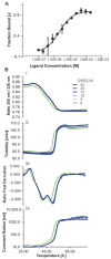
Biophysical analyses of the Ku-DBi 3392 interaction with Ku70–Ku80. (A) Interaction between Ku70–Ku80 with 3392 measured by MST (Microscale Thermophoresis) with a Kd of 2.1 ± 0.3 µM. The estimated bound fraction is plotted as a function of ligand concentration. (B) Impact of ligand 3392 on the thermostability and aggregation propensity of Ku70–Ku80: (i) ratio of intrinsic fluorescence at 350 nm divided by 330 nm, (ii) turbidity measurement, (iii) first derivative of the ratio, (iv) cumulant radius measured by DLS as a function of temperature.
Figure 3
Figure 3
Single-agent Ku-DBi activity of 3392 in comparison with NU-7441 in NSCLC cells. The indicated cell lines (A) H1299, (B) A549, (C) H460 and (D) H23 cells were plated and treated with the indicated agent for 48 h, and cell viability was determined using CCK-8 assays as described in Materials and Methods. Data represent the mean ± SEM of triplicate determinations.
Figure 4
Figure 4
Ku–DNA binding inhibition enhances cellular sensitivity to IR in NSCLC models. (A) Isobologram analysis for 3392 and bleomycin combination in H460 cells and response matrix for inhibition. (B) Surface plot for HSA and Bliss independence–additive models for synergy assessment. Data were analyzed using the Synergy finder tool v3.0 (https://synergyfinder.fimm.fi, accessed on June–July 2024). (C,D) Analyses of 3392 in combination with IR in (C) A549 and (D) H23 cells. Tukey’s multiple comparisons test, simple effects within rows. **** p< 0.0001, *** p< 0.001, ** p< 0.005, * p< 0.05.
Figure 5
Figure 5
Single-agent and combination cytotoxic activities of Ku-DBi 3392 in comparison with NU-7441 in TNBC cell lines. (A,B) The indicated cell lines were plated and treated with increasing concentrations of the indicated single agents, and cellular viability was determined by CCK-8 assay. (C,D) Bleomycin-sensitization activity was determined for the Ku-DBi and NU-7441 in the indicated cell lines. Cells were plated and treated as described in Materials and Methods and processed as described above. Data are presented as the mean ± SEM of triplicate determinations.
Figure 6
Figure 6
Impact of Ku-DBi in combination with IR on cell proliferation and early DDR events in NSCLC A549 CDX models. (A) Experimental design for combination Ku-DBi and IR treatment of NSCLC in vivo. (B) Western blot analysis from CDX tumor extracts assessing DNA-PKcs and γ-H2AX. (C,D) Quantification of the protein expression data presented in Panel B. (E) Representative 20X images of Ki-67 images, Sb: 100 mm. The insets show whole tumor sections for each treatment, Sb: 3 mm. Images were acquired using the Aperio ScanScope CS system. (F) Western blot detection of Ki-67 from the tumor tissue extracts. (G) Quantification of the data presented in panel F. Data are shown as mean ± SEM (vehicle n = 3, 3392 n = 3, IR n = 3, combination n = 4). Statistical analysis was performed using Fisher’s least significant difference test of the individual comparisons. Significant differences are indicated by * p < 0.05, ** p < 0.01, *** p < 0.001; ns: not significant. The original Western blot membranes can be found in Supplementary File S1.

References

    1. Blackford A.N., Jackson S.P. ATM, ATR, and DNA-PK: The Trinity at the Heart of the DNA Damage Response. Mol. Cell. 2017;66:801–817. doi: 10.1016/j.molcel.2017.05.015. - DOI - PubMed
    1. Jeggo P.A., Geuting V., Lobrich M. The role of homologous recombination in radiation-induced double-strand break repair. Radiother. Oncol. 2011;101:7–12. doi: 10.1016/j.radonc.2011.06.019. - DOI - PubMed
    1. Novotny J.P., Marino-Enriquez A., Fletcher J.A. Targeting DNA-PK. Cancer Treat. Res. 2023;186:299–312. - PMC - PubMed
    1. Pawelczak K.S., Gavande N.S., VanderVere-Carozza P.S., Turchi J.J. Modulating DNA Repair Pathways to Improve Precision Genome Engineering. ACS Chem. Biol. 2018;13:389–396. doi: 10.1021/acschembio.7b00777. - DOI - PubMed
    1. Gavande N.S., VanderVere-Carozza P.S., Pawelczak K.S., Mendoza-Munoz P., Vernon T.L., Hanakahi L.A., Summerlin M., Dynlacht J.R., Farmer A.H., Sears C.R., et al. Discovery and development of novel DNA-PK inhibitors by targeting the unique Ku-DNA interaction. Nucleic Acids Res. 2020;48:11536–11550. doi: 10.1093/nar/gkaa934. - DOI - PMC - PubMed

LinkOut - more resources