. 2024 Oct 8;17(19):4916.
doi: 10.3390/ma17194916.

In Vitro Investigation Using a New Biomechanical Force-Torque Analysis System: Comparison of Conventional and CAD/CAM-Fixed Orthodontic Retainers

Affiliations

In Vitro Investigation Using a New Biomechanical Force-Torque Analysis System: Comparison of Conventional and CAD/CAM-Fixed Orthodontic Retainers

Francesca Thaden et al. Materials (Basel). .

Abstract

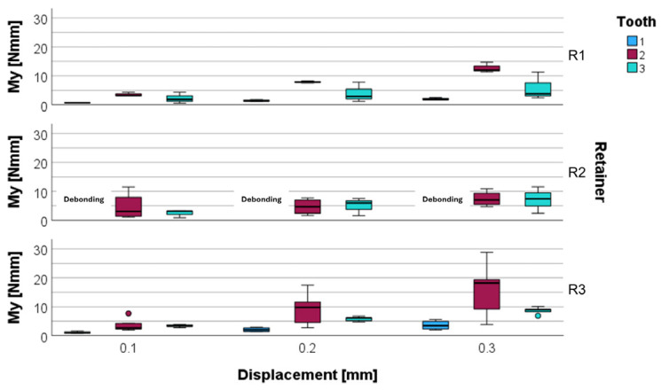
(1) Background: After more than a decade since their first description, Inadvertent Tooth Movements (ITMs) remain an adverse effect of orthodontic retainers without a clear etiology. To further investigate the link between ITMs and the mechanical properties of different retainers, the response upon vertical loading was compared in three retainer types (two stainless steel and one nickel-titanium). The influence of different reference teeth was also considered. (2) Methods: Three retainers (R1, R2, R3) were tested in a newly developed biomechanical analysis system (FRANS). They were bonded to 3D-printed models of the lower anterior jaw and vertically displaced up to 0.3 mm. Developing forces and moments were recorded at the center of force. (3) Results: The vertical displacement caused vertical forces (Fz) and labiolingual moments (My) to arise. These were highest in the lateral incisors (up to 2.35 ± 0.59 N and 9.27 ± 5.86 Nmm for R1; 1.69 ± 1.06 N and 7.42 ± 2.65 Nmm for R2; 3.28 ± 1.73 N and 15.91 ± 9.71 Nmm for R3) for all analyzed retainers and with the R3 retainer for all analyzed reference teeth, while the lowest Fz and My values were recorded with the R1 retainer. (4) Conclusions: Displacements of 0.2 mm and larger provided forces and moments which could be sufficient to cause unwanted torque movements, such as ITMs, in all analyzed retainers. Clinicians must be mindful of these risks and perform post-treatment checkups on patients with retainers of all materials.

Keywords: biomechanical phenomena; force; orthodontic retainer adverse effects; orthodontics; retainer; torque.


Conflict of interest statement

The authors declare no conflicts of interest.

Figures

Figure 1
Figure 1
The FRANS setup with all its components. [A] Digital Displacement Measuring Device; [B] micrometer screw gauge and dial gauge for vertical displacement; [C] springs to limit uncontrolled vertical movement; [D] Force/Torque sensor; [E] connector piece for force/moment transmission between sensor and reference teeth; [F] measurement table representing the lower jaw, with placement slots for the experimental setup representing the alveolar sockets; [G] screw gauge for the sagittal displacement of the measuring table. In this instance, the R2 retainer with the lateral incisors as reference teeth is being analyzed.
Figure 2
Figure 2
The defined coordinate system for the third and fourth quadrant, viewed from the lingual aspect. In both quadrants, the positive Y-axis indicates a mesial movement and the positive Z-axis indicates an intrusion. In the left quadrant, a positive X-axis indicates a labial movement; conversely, in the right quadrant, a positive X-axis indicates a lingual movement. Due to technical reasons, it was not possible to apply the Tweed convention to this coordinate system.
Figure 3
Figure 3
Measured force Fz [N] (left) and torque My [Nmm] (right) for all retainer types R1 (A,B), R2 (C,D), and R3 (E,F), exemplarily shown at the canines. The different measurements (a–e) are distinguished by color. While the curves for R3 (E,F) appear more homogenous, there are noticeable differences between measurements concerning R1 and R2.
Figure 4
Figure 4
Vertical force Fz for different retainer materials (R1, R2, R3) and different reference teeth (central incisor 1, lateral incisor 2, and canine 3) at a displacement of 0.1 mm, 0.2 mm, and 0.3 mm. The force Fz was measured at the center of force in N.
Figure 5
Figure 5
Labiolingual torque My for different retainer materials (R1, R2, R3) and different reference teeth (central incisor 1, lateral incisor 2, and canine 3) at a displacement of 0.1 mm, 0.2 mm, and 0.3 mm. The moment My was measured at the center of force in Nmm.
Figure 6
Figure 6
Biomechanical situation of incisal loading during mastication in the presence of a retainer. The retainer serves as center of rotation (CR). The effect is expected to be more pronounced when a twisted retainer wire is used, as the twisted wires are assumed to be less resistant to torsional moments such as My.
Figure 7
Figure 7
The lower anterior segment during vertical mastication loading causes a force in the negative Fz direction. Arrows indicating the vertical force-components on individual teeth (33, 32, 31, 41, 42, 43 according to the FDI notation). It becomes evident that the loaded canines (33 and 43) are only supported on one side whereas the lateral incisors (32 and 42) have support from the central incisors as well as from the canines. If the load is applied to the central incisors (31 and 41), the biomechanical support from the neighboring teeth is in between the two previously mentioned cases.

References

    1. Littlewood S.J. Evidence-based retention: Where are we now? Semin. Orthod. 2017;23:229–236. doi: 10.1053/j.sodo.2016.12.010. - DOI
    1. Melrose C., Millett D. Toward a perspective on orthodontic retention? Am. J. Orthod. Dentofac. Orthop. 1998;113:507–514. doi: 10.1016/S0889-5406(98)70261-6. - DOI - PubMed
    1. Reitan K. Clinical and histologic observations on tooth movement during and after orthodontic treatment. Am. J. Orthod. 1967;53:721–745. doi: 10.1016/0002-9416(67)90118-2. - DOI - PubMed
    1. Yu Y., Sun J., Lai W., Wu T., Koshy S., Shi Z. Interventions for managing relapse of the lower front teeth after orthodontic treatment. Cochrane Database Syst. Rev. 2013;2013:CD008734. doi: 10.1002/14651858.CD008734.pub2. - DOI - PMC - PubMed
    1. Kramer A., Sjostrom M., Hallman M., Feldmann I. Vacuum-formed retainer versus bonded retainer for dental stabilization in the mandible-a randomized controlled trial. Part I: Retentive capacity 6 and 18 months after orthodontic treatment. Eur. J. Orthod. 2020;42:551–558. doi: 10.1093/ejo/cjz072. - DOI - PubMed

LinkOut - more resources