Review
. 2024 Oct;7(10):e70016.
doi: 10.1002/cnr2.70016.

Prostate Cancer: A Review of Genetics, Current Biomarkers and Personalised Treatments

Affiliations
Review

Prostate Cancer: A Review of Genetics, Current Biomarkers and Personalised Treatments

Trevor K Wilson et al. Cancer Rep (Hoboken). 2024 Oct.

Abstract

Background: Prostate cancer is the second leading cause of cancer deaths in men, second only to lung cancer. Despite this, diagnosis and prognosis methods remain limited, with effective treatments being few and far between. Traditionally, prostate cancer is initially tested for through a prostate serum antigen (PSA) test and a digital rectum examination (DRE), followed by confirmation through an invasive prostate biopsy. The DRE and biopsy are uncomfortable for the patient, so less invasive, accurate diagnostic tools are needed. Current diagnostic tools, along with genes that hold possible biomarker uses in diagnosis, prognosis and indications for personalised treatment plans, were reviewed in this article.

Recent findings: Several genes from multiple families have been identified as possible biomarkers for disease, including those from the MYC and ETS families, as well as several tumour suppressor genes, Androgen Receptor signalling genes and DNA repair genes. There have also been advances in diagnostic tools, including MRI-targeted and liquid biopsies. Several personalised treatments have been developed over the years, including those that target metabolism-driven prostate cancer or those that target inflammation-driven cancer.

Conclusion: Several advances have been made in prostate cancer diagnosis and treatment, but the disease still grows year by year, leading to more and more deaths annually. This calls for even more research into this disease, allowing for better diagnosis and treatment methods and a better chance of patient survival.

Keywords: biomarkers; genes; personalised treatment; prostate cancer.


Conflict of interest statement

The authors declare no conflicts of interest.

Figures

FIGURE 1
FIGURE 1
Process of malignant transformation within prostate cells. The figure is taken from Rybak, Bristow and Kapoor [10] (Open License CC BY).
FIGURE 2
FIGURE 2
Mortality (red) and Age Standardised Incidence (blue) rates of common cancers in men, 2020. As can be seen, prostate cancer is the second leading cancer in terms of incidence, behind lung cancer, while being the sixth highest cancer in terms of mortality, falling behind lung, colorectum, stomach, liver and oesophageal cancer. Image Source: [13].
FIGURE 3
FIGURE 3
Map showing the worldwide ASR incidence rates for Prostate cancer based on 2020 statistics. Image Source: [13]. More developed countries have higher incidence rates than less developed countries, most likely due to more testing being conducted in these developed countries, leading to more cases being identified.
FIGURE 4
FIGURE 4
Graph showing the inverse relationship between prostate cancer mortality and incidence rates among continental populations. The regions with higher incidence rates tend to have a lower mortality rate. Data based on GLOBOCAN [13] statistics.
FIGURE 5
FIGURE 5
ASR incidence (blue) and Mortality (red) rates of the world's average and South Africa's population. Image Source: [13].
FIGURE 6
FIGURE 6
Progression of prostate cancer over time, showing the different general therapies used and at which stages the cancer becomes castration‐resistant, metastatic and symptomatic. Created using www.BioRender.com (2023).
FIGURE 7
FIGURE 7
Transverse section of the prostate indicating the positions from which the 12 cores are taken: 1–6 are the traditional sextant, 7–10 come from the peripheral zone and 11 and 12 come from the transition zone. Image Source: [81] (Open License CC BY).
FIGURE 8
FIGURE 8
Diagrammatic representation of the functions of the PTEN tumour suppressor, showing the role it plays in regulating the conversion of PIP3 to PIP2 and preventing the phosphorylation of AKT, thereby preventing irregular mTOR activation and dysregulating cellular functions. (A) PTEN regulates cell migration through protein substrate dephosphorylation; (B) PTEN inhibits the PI3K/Akt signalling cascade through PIP3 dephosphorylation to PIP2; (C) PTEN interacts with TP53, resulting in cell cycle arrest through increased TP53 stability; (D) PTEN preserve chromosome stability through interaction with the centromere. Image Source: [113] (Open License CC BY).
FIGURE 9
FIGURE 9
Process of prostate cancer cell proliferation through the dependence on testosterone (Tst) and its conversion to dihydrotestosterone (DHT), which binds with the AR, where it then moves to the nucleus, causing cell cycle targets transcription, resulting in cancer cell proliferation. Image created using BioRender (www.biorender.com). Inspiration from [118].
FIGURE 10
FIGURE 10
Diagram showing the process of DNA repair through homologous recombination controlled by multiple DNA repair proteins. Image Source: [124] (Open License CC BY).
FIGURE 11
FIGURE 11
Activation Process of the CHK2 protein. Firstly, as DNA damage is identified, ATM triggers the phosphorylation of the SQ/TQ cluster domain (SCD) of CHK2, leading to dimerisation and autophosphorylation of the kinase domain, triggering conformational changes resulting in dissociation and activation of monomer CHK2 proteins. Image created using BioRender (www.biorender.com).
FIGURE 12
FIGURE 12
Chemical structure of several drugs used to treat prostate cancer. (A) clioquinol; (B) quinone‐glucose conjugate created through conjugation of a glucose molecule and 1,4‐naphthoquinone; (C) etomoxir; (D) Enzalutamide (4‐[3‐[4‐cyano‐3‐(trifluoromethyl)phenyl]‐5,5 dimethyl‐4‐oxo‐2‐sulfanylideneimidazolidin‐1‐yl]‐2‐fluoro‐N‐methylbenzamide). Image Source: [190, 191, 192, 193] (Open License CC BY & Public Domain).
FIGURE 13
FIGURE 13
Haematoxylin and Eosin (HE) stain images of prostate tumours before and after treatment with AZ5069, showing tumour suppression and increased normalised prostate cells after treatment. Image Source: [212] (Open License CC BY‐NC‐ND).
FIGURE 14
FIGURE 14
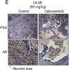
Cabozantinib treatment results in decreased AR and PSA immunoreactivity, with necrotic areas in the tumours after treatment. Image Source: [219] (Open License CC BY).
FIGURE 15
FIGURE 15
Prostate Cancer biopsy images showing the difference in myeloid cell counts before and after treatment with AZD5069 and enzalutamide. Scale bar = 100 μm. Image Source: [226] (Open License CC BY).
FIGURE 16
FIGURE 16
Diagram showing the different CYP enzymes involved in prostate cancer. CYP2R1, CYP27A1 and CYP27B1 are all downregulated in prostate cancer, while CYP24A1 is upregulated. Image created using www.BioRender.com (2024).
FIGURE 17
FIGURE 17
Diagram showing the effects of increased and decreased expression of the CYP3A and CYP2B6 enzymes in prostate cancer progression. Image created using BioRender (www.BioRender.com). Image Information: [230].

References

    1. Giona S., “The Epidemiology of Prostate Cancer,” in Prostate Cancer, eds. Simon M., Bott R. J., and Ng M. K. L. (Brisbane, Australia: Exon Publications, 2021), 1–16. - PubMed
    1. Pernar C. H., Ebot E. M., Wilson K. M., and Mucci L. A., “The Epidemiology of Prostate Cancer,” Cold Spring Harbor Perspectives in Medicine 8, no. 12 (2018): 1–19. - PMC - PubMed
    1. Shenoy D., Packianathan S., Chen A. M., et al., “Do African‐American Men Need Separate Prostate Cancer Screening Guidelines?,” BMC Urology 16, no. 1 (2016): 1–6. - PMC - PubMed
    1. Catalona W. J., “Prostate Cancer Screening,” Medical Clinics of North America 102, no. 2 (2018): 199–214. - PMC - PubMed
    1. Duffy M. J., “Biomarkers for Prostate Cancer: Prostate‐Specific Antigen and Beyond,” Clinical Chemistry and Laboratory Medicine (CCLM) 58, no. 3 (2020): 326–339. - PubMed

Substances