. 2024 Oct 1:4:1322851.
doi: 10.3389/fradi.2024.1322851. eCollection 2024.

Seven-tesla magnetic resonance imaging of the nervus terminalis, olfactory tracts, and olfactory bulbs in COVID-19 patients with anosmia and hypogeusia

Affiliations

Seven-tesla magnetic resonance imaging of the nervus terminalis, olfactory tracts, and olfactory bulbs in COVID-19 patients with anosmia and hypogeusia

Claudia F E Kirsch et al. Front Radiol. .

Abstract

Introduction: Linking olfactory epithelium to the central nervous system are cranial nerve 1, the olfactory nerve, and cranial nerve "0," and the nervus terminalis (NT). Since there is minimal expression of angiotensin-converting enzyme-2 (ACE-2) in the olfactory nerve, it is unclear how SARS-CoV-2 causes anosmia (loss of smell) and hypogeusia (reduction of taste). In animal models, NT expresses ACE-2 receptors, suggesting a possible SARS-CoV-2 viral entry site in humans. The purpose of this study was to determine whether ultra-high-field 7 T magnetic resonance imaging (MRI) could visualize the NT, olfactory bulbs (OB), and olfactory tract (OT) in healthy controls and COVID-19 anosmia or hypogeusia and to qualitatively assess for volume loss and T2 alterations.

Methods: In this study, 7 T MRI was used to evaluate the brain and olfactory regions in 45 COVID-19 patients and 29 healthy controls. Neuroimaging was qualitatively assessed by four board-certified neuroradiologists who were blinded to outcome assignments: for the presence or absence of NT; for OB, OT, and brain volume loss; and altered T2 signal, white matter T2 hyperintensities, microhemorrhages, enlarged perivascular spaces, and brainstem involvement.

Results: NT was identifiable in all COVID-19 patients and controls. T2 hyperintensity in the NT, OB, and OT in COVID-19 patients with anosmia or hypogeusia was statistically significant compared to controls and COVID-19 patients without anosmia or hypogeusia.

Discussion: On 7 T MRI, NT was radiographically identifiable, adjacent to OB and OT. In COVID-19 anosmia and hypogeusia, T2 hyperintensity of NT, OB, and OT was statistically significant compared to COVID-19 patients without anosmia or hypogeusia and controls. The NT may be a potential entry site for SARs-CoV-2 and may play a role in the pathophysiology of COVID-19 anosmia.

Keywords: angiotensin-converting enzyme 2 (ACE-2) receptor; hypothalamus; immune response; magnetic resonance imaging (MRI); nervus terminalis (NT); olfactory tract.


Conflict of interest statement

The authors declare that the research was conducted in the absence of any commercial or financial relationships that could be construed as a potential conflict of interest. The authors declared that they were an editorial board member of Frontiers at the time of submission. This had no impact on the peer review process and the final decision.

Figures

Figure 1
Figure 1
(A,B) Anatomy of the nasal cavity, olfactory apparatus including the olfactory bulbs (OBs), olfactory tracts (OTs), and nerves (yellow circles), nervus terminals (NT) (red oval, red lines, red arrows), nasal mucosa (tea-colored circle), and ethmoid bone and foramina (beige circle) (C. Kirsch, Primal pictures 3D anatomy). With permission © Pharma Intelligence UK (trading as Primal Pictures), 2024. www.primalpictures.com www.anatomy.tv.
Figure 2
Figure 2
UHF 7-T MRI T2-weighted (A,B) coronal and (C) axial scans of a 38-year-old female healthy control. In (C), the orbital globe, pink dual arrow, measures 2.5 cm anterior–posterior for scale. (D) UHF 7-T MR T2-weighted coronal images of a 33- year-old female healthy control. Yellow arrows point to olfactory bulbs in (A,D) and olfactory tracts, red arrows point to the NT, blue arrowheads point to vessels with flow voids, and curved green arrows point to the olfactory sulcus. The depth of the olfactory sulcus in the coronal plane measures between 6.5 and 7.5 mm.
Figure 3
Figure 3
UHF 7-T MRI T2-weighted (A–C) coronal and (D,E) axial scans of a 43-year-old female with anosmia and hypogeusia during and persisting after recovery from COVID-19. Yellow arrows point to olfactory bulbs, red arrows point to the NT, blue arrowheads point to vessels with flow voids, and curved green arrows point to the olfactory sulcus. There is an increased T2 signal in the bilateral olfactory bulbs, with the left greater than right olfactory tracts and NT, as well as left volume loss greater than right volume loss in the left olfactory bulb and tract, with an asymmetric widening of the left olfactory sulcus.
Figure 4
Figure 4
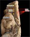
Autopsy brain specimen, yellow arrowhead points to olfactory bulb (OB), yellow arrows point to olfactory tract (OT), red arrows point to nervus terminalis (NT) the thin unmyelinated fibers adjacent to the OB and OT.

References

    1. Lan J, Ge J, Yu J, Shan S, Zhou H, Fan S, et al. Structure of the SARS-CoV-2 spike receptor-binding domain bound to the ACE2 receptor. Nature. (2020) 581(7807):215–20. 10.1038/s41586-020-2180-52 - DOI - PubMed
    1. Borkotoky S, Dey D, Hazarika Z. Interactions of angiotensin-converting enzyme-2 (ACE2) and SARS-CoV-2 spike receptor-binding domain (RBD): a structural perspective. Mol Biol Rep. (2023) 50(3):2713–21. 10.1007/s11033-022-08193-4 - DOI - PMC - PubMed
    1. Najjar S, Najjar A, Chong DJ, Pramanik BK, Kirsch C, Kuzniecky RI, et al. Central nervous system complications associated with SARS-CoV-2 infection: integrative concepts of pathophysiology and case reports. J Neuroinflammation. (2020) 17(1):231. 10.1186/s12974-020-01896-0 - DOI - PMC - PubMed
    1. Hoffmann M, Kleine-Weber H, Schroeder S, Krüger N, Herrler T, Erichsen S, et al. SARS-CoV-2 cell entry depends on ACE2 and TMPRSS2 and is blocked by a clinically proven protease inhibitor. Cell. (2020) 181(2):271–80.e8. 10.1016/j.cell.2020.02.052 - DOI - PMC - PubMed
    1. Meinhardt J, Streit S, Dittmayer C, Manitius RV, Radbruch H, Heppner FL. The neurobiology of SARS-CoV-2 infection. Nat Rev Neurosci. (2024) 25(1):30–42. 10.1038/s41583-023-00769-8 - DOI - PubMed

LinkOut - more resources