. 2024 Oct 1:15:1439448.
doi: 10.3389/fphar.2024.1439448. eCollection 2024.

Hemoglobin α-derived peptides VD-hemopressin (α) and RVD-hemopressin (α) are involved in electroacupuncture inhibition of chronic pain

Affiliations

Hemoglobin α-derived peptides VD-hemopressin (α) and RVD-hemopressin (α) are involved in electroacupuncture inhibition of chronic pain

Xiaocui Yuan et al. Front Pharmacol. .

Abstract

Introduction: Knee osteoarthritis (KOA) is a chronic degenerative bone metabolic disease that primarily affects older adults, leading to chronic pain and disability that affect patients' daily activities. Electroacupuncture (EA) is a commonly used method for the treatment of chronic pain in clinical practice. Previous studies indicate that the endocannabinoid system is involved in EA analgesia, but whether endocannabinopeptide VD-hemopressin (α) and RVD-hemopressin (α) derived from hemoglobin chains are involved in EA analgesia is unclear.

Methods: RNA-seq technology was used to screen which genes involved in EA analgesia. The expression of hemoglobin α chain and 26S proteasome were determined by Western blotting. The level of VD-hemopressin (α) and RVD-hemopressin (α) were measured by UPLC-MS/MS. Microinjection VD-Hemopressin (α), RVD-Hemopressin (α) and 26S proteasome inhibitor MG-132 into vlPAG, then observe mechanical and thermal pain thresholds.

Results: Therefore, we used RNA-seq to obtain differentially expressed genes Hba-a1 and Hba-a2 involved in EA analgesia in the periaqueductal gray (PAG), which were translated into the hemoglobin α chain. EA significantly increased the expression of the hemoglobin α chain and the level of hemopressin (α) and RVD-hemopressin (α). Microinjection of VD-hemopressin (α) and RVD-hemopressin (α) into the ventrolateral periaqueductal gray (vlPAG) mimicked the analgesic effect of EA, while CB1 receptor antagonist AM251 reversed this effect. EA significantly increased the expression of 26S proteasome in KOA mice. Microinjection of 26S proteasome inhibitor MG132 before EA prevented both the anti-allodynic effect and upregulation of the concentration of RVD-hemopressin (α) by EA treatment and upregulated the expression of the hemoglobin α chain.

Discussion: Our data suggest that EA upregulated the concentration of VD-hemopressin (α) and RVD-hemopressin (α) through enhancement of the hemoglobin α chain degradation by 26S proteasome in the PAG, then activated the CB1 receptor, thereby exerting inhibition of chronic pain in a mouse model of KOA. These results provide new insights into the EA analgesic mechanisms and reveal possible targets for EA treatment of chronic pain.

Keywords: 26S proteasome; Chronic pain; Electroacupuncture analgesia; Knee osteoarthritis (KOA); RVD-hemopressin (α); VD-hemopressin (α).


Conflict of interest statement

The authors declare that the research was conducted in the absence of any commercial or financial relationships that could be construed as a potential conflict of interest.

Figures

FIGURE 1
FIGURE 1
Screening differentially expressed genes (DEGs) Hba-a1 and Hba-a2 involved in EA analgesia in the periaqueductal gray. (A) DEG statistics. The X-axis represents pairwise comparisons, and the Y-axis shows the number of screened DEGs. The blue bars denote downregulated genes, and the orange bars denote the upregulated ones. (B) Heatmap of DEGs in the KOA, EA, and control groups. (C) Summary data show the effect of KOA and EA on the level of the Hba-a1 in PAG tissues. (D) Summary data show the effect of KOA and EA on the level of the Hba-a2 in PAG tissues. Data are expressed as means ± SEM (n = 3 mice in each group). *p < 0.05.
FIGURE 2
FIGURE 2
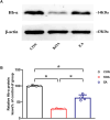
Quantitative analysis of the protein level of hemoglobin α-chain in the PAG tissues. (A) The representative gel image shows the protein level of the hemoglobin α-chain in PAG tissues obtained from the control (CON), KOA, and KOA treated with EA groups. β-actin was used as a loading control. The protein band at 14 kDa corresponds to the hemoglobin α-chain. (B) Summary data show the effect of KOA and EA on the protein level of the hemoglobin α-chain in PAG tissues. Data are expressed as means ± SEM (n = 4 mice in each group). *p < 0.05.
FIGURE 3
FIGURE 3
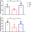
Quantitative analysis of the VD-hemopressin (α) and RVD-hemopressin (α) concentrations in the PAG. (A) Summary data show the effect of KOA and EA on the concentration of VD-hemopressin (α) in the PAG. (B) Summary data show the effect of KOA and EA on the concentration of RVD-hemopressin (α) in the PAG. Data are expressed as means ± SEM (n = 4 mice in each group). *p < 0.05.
FIGURE 4
FIGURE 4
Time course of the effect of VD-hemopressin (α) and RVD-hemopressin (α) on pain hypersensitivity in KOA mice. (A, B) Time course of tactile threshold in response to von Frey filaments or a noxious heat stimulus in CON, KOA + saline, EA, and 10 nmol VD-hemopressin (α) mice. (C, D) Time course of the effect of different concentrations of RVD-hemopressin (α) on tactile and thermal withdrawal thresholds of KOA mice. VD-hemopressin (α) or RVD-hemopressin (α) was microinjected into the vlPAG, or the mice received EA stimulation starting from 18 days after MIA injection, once every other day for five times, as indicated by the black arrow. Data are expressed as means ± SEM (n = 8–11 mice in each group). *p < 0.05, compared with the KOA + Saline group; #p < 0.05, compared with the 1 nmol RVD-hemopressin (α) group; + p < 0.05, compared with the 2 nmol RVD-hemopressin (α) group.
FIGURE 5
FIGURE 5
Effects of the CB1 receptor antagonist AM251 on VD-hemopressin (α) and RVD-hemopressin (α) analgesia. (A, B) Time course of the effect of the CB1 receptor antagonist AM251 on tactile and thermal withdrawal thresholds of VD-hemopressin (α) mice. (C, D) Time course of the effect of the CB1 receptor antagonist AM251 on tactile and thermal withdrawal thresholds of RVD-hemopressin (α) mice. The CB1 receptor antagonist AM251 was microinjected into the vlPAG 10 min before VD-hemopressin (α) or RVD-hemopressin (α) starting from 18 days after MIA injection, once every other day for five times, as indicated by the black arrow. *p < 0.05; ***p < 0.001, compared with KOA group; ###p < 0.001, compared with the 10 nmol VD-hemopressin (α) group; +++ p < 0.001, compared with the 5-nmol RVD-hemopressin (α) group. Data are expressed as means ± SEM (n = 5–11 mice in each group).
FIGURE 6
FIGURE 6
Quantitative analysis of the protein level of 26S proteasome (PSMD2) in the PAG tissues. (A) The representative gel image shows the protein level of the PSMD2 in PAG tissues obtained from control (CON), KOA, and KOA treated with EA. β-actin was used as a loading control. The protein band at 100 kDa corresponds to the PSMD2. (B) Summary data show the effect of KOA and EA on the protein level of the PSMD2 in PAG tissues. Data are expressed as means ± SEM (n = 4 mice in each group). ***p < 0.001.
FIGURE 7
FIGURE 7
Effects of the 26S proteasome inhibitor MG132 on EA analgesia and RVD-hemopressin (α) concentration in the PAG. (A, B) Time course of the effect of the 26S proteasome inhibitor MG132 on tactile and thermal withdrawal thresholds of EA mice. EA was administered for 30 min, once every other day for 4 weeks, starting from 2 days after the MIA injection. The 26S proteasome inhibitor MG132 (4 μg) was microinjected into the vlPAG 30 min before EA starting from 18 days after MIA injection, once every other day for five times, as indicated by the black arrow. Data are expressed as means ± SEM (n = 8–11 mice in each group). *p < 0.05, compared with the KOA group; #p < 0.05, compared with the 10% DMSO + EA group. (C) Summary data show the concentration of RVD-hemopressin (α) in the PAG. Data are expressed as means ± SEM (n = 4–6 mice in each group). *p < 0.001.
FIGURE 8
FIGURE 8
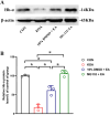
Effects of the 26S proteasome inhibitor MG132 on hemoglobin α-chain expression in the PAG. (A) The representative gel image shows the protein level of hemoglobin α-chain in PAG tissues obtained from CON, KOA, 10% DMSO + EA, and MG132 + EA groups. β-actin was used as a loading control. The protein band at 14 kDa corresponds to the hemoglobin α-chain. (B) Summary data show the effect of 26S proteasome inhibitor MG132 on the protein level of the hemoglobin α-chain in PAG tissues. EA was administered for 30 min, once every other day for 4 weeks, starting from 2 days after MIA injection. The 26S proteasome inhibitor MG132 (4 μg) was microinjected into the vlPAG 30 min before EA starting from 18 days after MIA injection, once every other day for five times. Data are expressed as means ± SEM (n = 3 mice in each group). *p < 0.05.

References

    1. Bauer M., Chicca A., Tamborrini M., Eisen D., Lerner R., Lutz B., et al. (2012). Identification and quantification of a new family of peptide endocannabinoids (Pepcans) showing negative allosteric modulation at CB1 receptors. J. Biol. Chem. 287 (44), 36944–36967. 10.1074/jbc.M112.382481 - DOI - PMC - PubMed
    1. Biagioli M., Pinto M., Cesselli D., Zaninello M., Lazarevic D., Roncaglia P., et al. (2009). Unexpected expression of alpha- and beta-globin in mesencephalic dopaminergic neurons and glial cells. Proc. Natl. Acad. Sci. U. S. A. 106 (36), 15454–15459. 10.1073/pnas.0813216106 - DOI - PMC - PubMed
    1. Chaplan S. R., Bach F. W., Pogrel J. W., Chung J. M., Yaksh T. L. (1994). Quantitative assessment of tactile allodynia in the rat paw. J. Neurosci. Methods 53 (1), 55–63. 10.1016/0165-0270(94)90144-9 - DOI - PubMed
    1. Chen L., Zhang J., Li F., Qiu Y., Wang L., Li Y. H., et al. (2009). Endogenous anandamide and cannabinoid receptor-2 contribute to electroacupuncture analgesia in rats. J. Pain 10 (7), 732–739. 10.1016/j.jpain.2008.12.012 - DOI - PubMed
    1. Chen Y., Zhou Y., Li X. C., Ma X., Mi W. L., Chu Y. X., et al. (2022). Neuronal GRK2 regulates microglial activation and contributes to electroacupuncture analgesia on inflammatory pain in mice. Biol. Res. 55 (1), 5. 10.1186/s40659-022-00374-6 - DOI - PMC - PubMed

LinkOut - more resources