. 2024 Oct 1;13(10):25.
doi: 10.1167/tvst.13.10.25.

Multi-Characteristic Opsin Therapy to Functionalize Retina, Attenuate Retinal Degeneration, and Restore Vision in Mouse Models of Retinitis Pigmentosa

Affiliations

Multi-Characteristic Opsin Therapy to Functionalize Retina, Attenuate Retinal Degeneration, and Restore Vision in Mouse Models of Retinitis Pigmentosa

Subrata Batabyal et al. Transl Vis Sci Technol. .

Abstract

Purpose: Retinal degeneration 1 and 10 (rd1 and rd10) mice are useful animal models of retinitis pigmentosa (RP) with rapidly and slowly progressive pathologies, respectively. Our study aims were to determine the effect of adeno-associated viral vector 2 (AAV2)-delivered multi-characteristic opsin (MCO-010; under the control of a metabotropic glutamate receptor-6 promoter enhancer) on the morphological and functional characteristics of vision in both rd1 and rd10 mice.

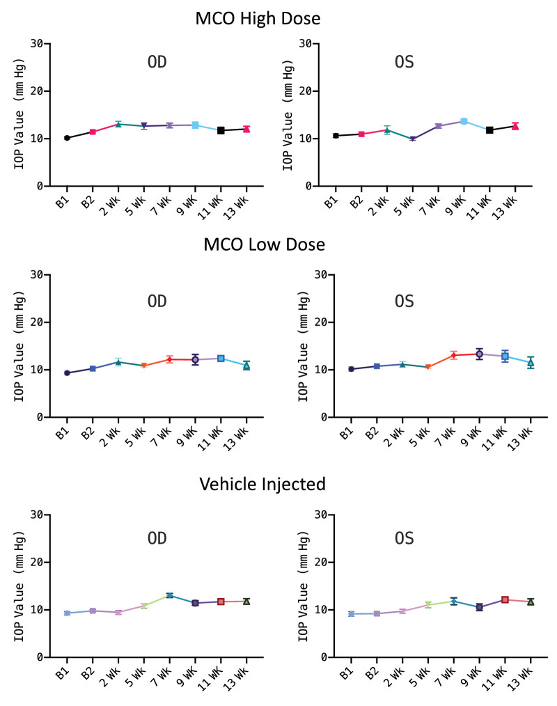
Methods: Various retinal measures of MCO-010 transduction and electrophysiological, behavioral, and other routine blood analyses were performed in the rd1 and/or rd10 mice after intravitreal injection of 1 µL of MCO-010 or AAV2 vehicle. Functional tests included electroretinogram, visually evoked potential, and behavior assay (optomotor and water maze). Retinal thickness, intraocular pressure, and plasma cytokine levels were also determined.

Results: Following intravitreal MCO-010 injection, approximately 80% of bipolar cells were transduced in the retina, and no alterations in retinal thickness were observed at 4 months post-injection. However, retinal thickness significantly decreased in control mice. MCO-010 treatment increased head movements and induced faster navigation of mice to the platform in a water-maze test. The MCO-010 gene therapy helped preserve visually evoked electrical response in the retina and visual cortex. No ocular toxicity, immunotoxicity, or phototoxicity was observed in the MCO-010-treated mice, even under chronic intense light conditions.

Conclusions: Intravitreal MCO-010 was well tolerated in rd1 and rd10 mice models of RP, and it appeared to attenuate retinal photoreceptor degeneration based on retinal structure and functional outcome measures.

Translational relevance: As reported here, optogenetic treatment of the inner retina attenuates further retinal degeneration in addition to photosensitizing higher order neurons, and this disease-modifying aspect should be evaluated in optogenetic clinical trials.


Conflict of interest statement

Disclosure: S. Batabyal, Nanoscope Therapeutics (I); S. Kim, Nanoscope Therapeutics (I); M. Carlson, Nanoscope Therapeutics (I); D. Narcisse, None; K. Tchedre, Nanoscope Therapeutics (I); A. Dibas, Nanoscope Therapeutics (I); N.A. Sharif, Nanoscope Therapeutics (I); S. Mohanty, Nanoscope Therapeutics (I), Nanoscope Technologies (I)

Figures

Figure 1.
Figure 1.
Intravitreal injection of MCO-010 led to selective expression in rd10 mouse retina. The retina slices were immunostained using primary antibodies (for mCherry and PKCα). (A) PKCα (green) shows the soma and axonal terminals of the bipolar cells. (B) Expression of MCO in bipolar cells visualized by an immunoassay for mCherry (red). (C) Zoomed-in region of the rectangle marked in (A) showing the expression of MCO in bipolar cells. (D) Quantification of percentage of mCherry-positive bipolar cells in MCO-010–injected and control groups. (E) Staining for PKCα in the not injected control mice. (F) Staining for mCherry. Average ± SD; *P < 0.0001.
Figure 2.
Figure 2.
Intravitreal injection of MCO-010 attenuated retina degeneration, as evaluated by SD-OCT and immunohistology. (A) Box plot of thickness of retina of rd mice before and after different doses of MCO-010 or AAV vehicle injection. Group AA included 3.4E8 gc/eye MCO-010; group BB included 1E9 vg/eye AAV2 (no transgene); and group CC included 3.4E6 gc/eye MCO-010. (B) Scatterplot showing comparisons of retinal thickness before and 4 months after injection in the different groups. (C) The mean difference of retinal thickness between baseline and intravitreally injected mice shown as a Gardner–Altman estimation plot. *P < 0.05 for group BB between baseline and 4 months after vehicle injection. There was no statistically significant difference observed in MCO-010–injected groups AA and CC. (D) MCO-010 treatment led to enhanced connectivity of the synaptic terminals of bipolar cells with RGCs measured by CtBP (red) staining. (E) CtBP immunostaining for control non-treated retina. Right panels of (D) and (E) show composites of CtBP with PKCα (green) and DAPI (blue). (F) Quantification of PKCα and CtBP fluorescence in MCO-treated and non-treated mice (mean ± SEM; *P < 0.05).
Figure 3.
Figure 3.
Longitudinal monitoring of optomotor responses of rd1 mice before and after injection of MCO-010 or AAV2 (vehicle control). (A) Number of head movements at 0.25 cpd during baseline and at 13-weeks post-injection for individual mice (n = 3 mice per group; mean ± SD). F, female; M, male. (B) Comparison of baseline-subtracted number of head movements at baseline and different time points after intravitreal injection of MCO-010 and AAV2 vehicle (n = 10 mice per group; mean ± SEM; *P < 0.05). The light intensity at the center of the chamber was 1 µW/mm2.
Figure 4.
Figure 4.
Visually guided dose-dependent behavioral improvement in young and old rd10 mice in radial-water-maze tests after MCO-010 injection. (A, B) Times to reach the platform from the center of the radial-arm water maze at baseline (B1, B2, and B3) and at post-injection evaluations performed at weeks 3 to 26 in 3-month-old mice intravitreally injected with MCO-010: (A) 3E9 gc/eye, (B) 3.4E8 gc/eye (n = 6 mice, 3 male and 3 female, per dose group). (C) Times to reach the platform from the center of the radial-arm water maze at baseline (B1 and B2) and post-injection evaluations at weeks 4 to 24 in 6-month-old mice intravitreally injected with MCO-010 (1.2E9 gc/eye; n = 6 female mice). LED light intensity was 7 µW/mm2 (mean ± SEM; *P < 0.05 between baseline and different time points). (D) Visually guided behavior in the water-maze test of 6-month-old rd10 mice improved after injection of MCO-010 as compared to AAV2 (vehicle control)–injected age-matched mice (n = 5 mice per group). The light intensity was 7 µW/mm2 (mean ± SEM; *P < 0.05).
Figure 5.
Figure 5.
Fast electrical responses of retina in MCO-010–injected rd10 mice observed using electrophysiological measurements. (A) Validation of the light-evoked fast electrophysiological responses in rd1 (negative control) and wild-type (positive control) mice. (B) Representative electrical responses of retinas before and 4 weeks after MCO-010 treatment of rd10 mice with and without light stimulation (1 ms, indicated by red bars). (C) Representative longitudinal measurement (baseline and 4 and 13 weeks post-injection) of fast electrical responses in MCO-010–injected OD and OS eyes of rd10 mice in response to 1-ms light pulses (3 cd·s/m2). The timing of the light stimulus is shown by the arrow.
Figure 6.
Figure 6.
VEPs in rd1 mice with and without intravitreal injections of MCO-010. (A) Representative VEPs of rd1 mice at a light intensity of 7 × 1012 photons/cm2/s, 8 months after AAV2 vehicle injection (1E9 vg/eye). (B) VEPs of rd1 mice 8 months after injection with MCO-010 (3.4E8 gc/eye) at a light intensity of 7 × 1012 photons/cm2/s. The downward arrow (at 0 ms) indicates the point of light stimulation. (C) Quantitative comparison of the amplitudes of negative peaks of VEPs in rd1 mice receiving intravitreal injections of MCO-010 or AAV control (mean ± SEM; n = 4 mice; *P < 0.05).
Figure 7.
Figure 7.
Intravitreal injection of MCO-010 did not increase the IOP of rd1 mice. Both eyes were injected with either AAV2 vehicle control or MCO-010. Low dose: 3.4E6 gc/eye (n = 5); high dose: 3.4E8 gc/eye (n = 10); vehicle (AAV2) dose: 1E9 vg/eye (n = 9). Left, OD; right, OS. Average ± SEM.
Figure 8.
Figure 8.
Intravitreal injection of MCO-010 in rd10 mice did not cause long-term photo- or immunotoxicity. Figure shows long-term viability of retinal bipolar cells after 4 months of chronic light exposure assessed by caspase assay. (A) Representative fluorescence images of retina stained with caspase-3 (red) overlaid with PKCα (green) for MCO-010–treated mouse retina, 4 months after 8-hr/day illumination of white light (intensity, 0.1 mW/mm2). (Top) OD eye and (bottom) non-treated contralateral OS eye. Scale bar: 50 µm. No caspase-3–positive apoptotic cells were observed in the INL of MCO-010–treated rd10 mice after 4 months of chronic light exposure (n = 4 mice). (B) Long-term immunotoxicity in plasma of MCO-010–injected rd10 mice. (Left) Longitudinal changes in IL-6 (pro-inflammatory marker) levels in the plasma of mice injected with MCO-010 (3.4E8 vg/eye) as compared to baseline values. (Right) Changes in IL-10 (anti-inflammatory marker) levels over the 6-month post-injection period (mean ± SD; n = 6 animals).

References

    1. Hartong DT, Berson EL, Dryja TP.. Retinitis pigmentosa. Lancet. 2006; 368: 1795–1809. - PubMed
    1. Sugawara T, Hagiwara A, Hiramatsu A, Ogata K, Mitamura Y, Yamamoto S.. Relationship between peripheral visual field loss and vision-related quality of life in patients with retinitis pigmentosa. Eye (Lond). 2010; 24: 535–539. - PubMed
    1. Daiger SP, Bowne SJ, Sullivan LS.. Perspective on genes and mutations causing retinitis pigmentosa. Arch Ophthalmol. 2007; 125: 151–158. - PMC - PubMed
    1. Mezer E, Babul-Hirji R, Wise R, et al. .. Attitudes regarding predictive testing for retinitis pigmentosa. Ophthalmic Genet. 2007; 28: 9–15. - PubMed
    1. Flannery JG, Farber DB, Bird AC, Bok D.. Degenerative changes in a retina affected with autosomal dominant retinitis pigmentosa. Invest Ophthalmol Vis Sci. 1989; 30: 191–211. - PubMed

Publication types

MeSH terms

Supplementary concepts