. 2024 Oct 16;20(10):e1012622.
doi: 10.1371/journal.ppat.1012622. eCollection 2024 Oct.

Swine NONO promotes IRF3-mediated antiviral immune response by Detecting PRRSV N protein

Affiliations

Swine NONO promotes IRF3-mediated antiviral immune response by Detecting PRRSV N protein

Dandan Jiang et al. PLoS Pathog. .

Abstract

Non-POU domain-containing octamer-binding protein (NONO) is a multi-functional nuclear protein which belongs to the Drosophila behavior/human splicing (DBHS) protein family. NONO is known to regulate multiple important biological processes including host antiviral immune response. However, whether NONO can inhibit porcine reproductive and respiratory syndrome virus (PRRSV) replication is less well understood. In this study, we demonstrated that swine NONO (sNONO) inhibited PRRSV replication, via increasing expression of IFN-β, whereas NONO knockdown or knockout in PAM-KNU cells was more susceptible to PRRSV infection. As an IRF3 positive regulation factor, NONO promoted IFN-β expression by enhancing activation of IRF3. During PRRSV infection, NONO further up-regulated IRF3-mediated IFN-β expression by interacting with PRRSV N protein. Mechanistically, NONO functioned as a scaffold protein to detect PRRSV N protein and formed N-NONO-IRF3 complex in the nucleus. Interestingly, it was found that the NONO protein reversed the inhibitory effect of PRRSV N protein on type I IFN signaling pathway. Taken together, our study provides a novel mechanism for NONO to increase the IRF3-mediated IFN-β activation by interacting with the viral N protein to inhibit PRRSV infection.


Conflict of interest statement

The authors have declared that no competing interests exist.

Figures

Fig 1
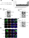
Fig 1. NONO inhibits PRRSV replication.
(A) PAM-KNU cells were transfected with 10 pmol of control siRNA (siCtrl) or siRNA targeting NONO (siNONO) for 24 h. The mRNA levels of NONO were detected by real-time PCR (n = 3 independent experiment, **p < 0.01, bar indicates mean). The protein levels of NONO were detected by Western blotting with anti-NONO antibody. The same blot was incubated with β-actin antibody as a protein loading control (n = 3 independent experiment, one representative experiment is shown). (B-D) PAM-KNU cells were transfected with 10 pmol of siNONO or siCtrl for 24 h and then infected with HP-PRRSV SY0608 strain at an MOI of 1 (B). PAM-KNU cells were transfected with 1 μg of pXJ41-sNONO or pXJ41 for 24 h and infected with HP-PRRSV SY0608 strain at an MOI of 1 (C). PAM-KNU and sNONO KO-PAM-KNU cells were infected with HP-PRRSV SY0608 strain at an MOI of 1 (D). Culture supernatants were collected at indicated times and viral RNA was extracted from culture supernatants to analyze by real-time PCR. Virus titers in culture supernatants were measured by microtitration infectivity assay and calculated TCID50 using the Reed-Muench method (n = 3 independent experiment, *p < 0.05, **p < 0.01, bar indicates mean). Whole-cell lysates were immunoblotted with anti-FLAG, anti-NONO, anti-N, or anti-ISG15 antibody. The same blot was incubated with β-actin antibody as a protein loading control (n = 3 independent experiment, one representative experiment is shown). The band intensities of N are shown as the relative protein expression levels, normalized with β-actin (n = 3 independent experiment, *p < 0.05, **p < 0.01, bar indicates mean).
Fig 2
Fig 2. NONO positively regulates IFN-β signaling pathway.
(A-C) PAM-KNU cells were transfected with 10 pmol of siNONO or siCtrl for 24 h and then treated with 1 μg/mL poly (I:C) for 6 h (A). PAM-KNU cells were transfected with 1 μg of pXJ41-sNONO or pXJ41 for 24 h and treated with 1 μg/mL poly (I:C) for 6 h (B). PAM-KNU and sNONO KO-PAM-KNU cells were treated with 1 μg/mL poly (I:C) for 6 h (C). Cells were harvested and total RNA was extracted using TRIzol reagent. The mRNA levels of IFN-β, ISG15 and Mx1 were analyzed by real-time PCR (n = 3 independent experiment, **p < 0.01, bar indicates mean).
Fig 3
Fig 3. NONO interacts with IRF3 and promotes IRF3-mediated IFN-β signaling pathway.
(A) PAM-KNU cells were transfected with 6 μg of pXJ41-sNONO or pXJ41 for 24 h. Then NONO complex was pulled down by immunoprecipitation with anti-FLAG antibody. The precipitation was subjected to SDS-PAGE and stained with Coomassie Blue, followed by analysis using protein affinity purification mass spectrometry (AP-MS). (B) Significant IRF3 enriched in NONO immunoprecipitates by quantitative AP-MS (n = 3 independent experiments). (C) The sequence alignment between swine and human NONO. (D) HEK-293T or PAM-KNU cells were cotransfected with 3 μg of pXJ41-sNONO and 3 μg of pXJ41-sIRF3 or pXJ41 for 24 h, followed by co-IP with anti-FLAG antibody and immunoblot analysis with anti-FLAG, anti-MYC or anti-IRF3 antibody. Whole-cell lysis (WCL) was subjected to Western blotting using anti-FLAG, anti-MYC, anti-IRF3, or anti-β-actin antibody (n = 3 independent experiment, one representative experiment is shown). (E) PAM-KNU cells were transfected with 0.5 μg of pXJ41-sNONO and 0.5 μg of pXJ41-sIRF3 or pXJ41 for 24 h. The cells were harvested and total RNA was extracted by TRIzol reagent. The mRNA level of IFN-β was analyzed by real-time PCR (n = 3 independent experiment, **p < 0.01, bar indicates mean). (F) PAM-KNU cells were transfected with 0.3 μg of pXJ41-sNONO and 0.3 μg of pXJ41-sIRF3 or pXJ41, along with 0.3 μg of pIFN-β-Luc and 0.03 μg of pRL-TK luciferase reporter for 24 h. IFN-β promoter activity was detected by a dual-luciferase reporter assay (n = 3 independent experiment, **p < 0.01, bar indicates mean). (G) HEK-293T cells were transfected with 0.3 μg of pXJ41-sNONO and 0.3 μg of pXJ41-hIRF3 or pXJ41, along with 0.3 μg of pIFN-β-Luc and 0.03 μg of pRL-TK luciferase reporter for 24 h. IFN-β promoter activity was detected by a dual-luciferase reporter assay (n = 3 independent experiment, **p < 0.01, bar indicates mean).
Fig 4
Fig 4. NONO interacts with PRRSV N protein.
(A) Schematic representation of LC-NONO and LN-PRRSV proteins. (B) HEK-293T cells were cotransfected with 0.5 μg of LC-NONO and 0.5 μg of LN-PRRSV constructs for 24 h. Cells were harvested and analyzed for recombination Renilla luciferase reporter activity by luciferase assay system (n = 3 independent experiment, **p < 0.01, bar indicates mean). (C) HEK-293T cells were cotransfected with 3 μg of pXJ41-sNONO and 3 μg of pXJ41-N or pXJ41 for 24 h. Cells were harvested and subjected to co-IP with anti-FLAG or anti-HA antibody. Immunocomplexes were analyzed by Western blotting using anti-HA or anti-FLAG antibody. WCL was also subjected to Western blotting using anti-HA, anti-FLAG, or anti-β-actin antibody (n = 3 independent experiment, one representative experiment is shown). (D) PAM-KNU cells were cotransfected with 0.25 μg of pXJ41-sNONO and 0.25 μg of pXJ41-N or pXJ41 for 18 h. Indirect immunofluorescence (IFA) was performed using anti-HA antibody (green), anti-FLAG antibody (red), or DAPI (blue). Scale bar, 10 μm. (E) PAM-KNU cells were transfected with 6 μg of pXJ41-sNONO or pXJ41 for 18 h and then infected with PRRSV at an MOI of 1 for 12 h. Cells were harvested and subjected to co-IP with anti-FLAG antibody. Immunocomplexes were analyzed by Western blotting using anti-N antibody. WCL was also subjected to Western blotting using anti-FLAG, anti-N, or anti-β-actin antibody (n = 3 independent experiment, one representative experiment is shown). (F) PAM-KNU cells were transfected with 0.25 μg of pXJ41-sNONO or pXJ41 for 18 h and infected with PRRSV at an MOI of 1 for 12 h. Indirect immunofluorescence (IFA) was performed using anti-N antibody (green), anti-FLAG antibody (red), or DAPI (blue). Scale bar, 10 μm.
Fig 5
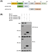
Fig 5. PRRSV N associates with N-terminal domain of NONO.
(A) Schematic representation of swine NONO structure. (B) HEK-293T cells were cotransfected with 3 μg of pXJ41-N and 3 μg of pXJ41-sNONO, pXJ41-sNONO-NTD or pXJ41-sNONO-CTD for 24 h. Cells were harvested and subjected to co-IP with anti-FLAG antibody. Immunocomplexes were analyzed by Western blotting using anti-HA antibody. WCL was also subjected to Western blotting using anti-HA, anti-FLAG, or anti-β-actin antibody (n = 3 independent experiment, one representative experiment is shown).
Fig 6
Fig 6. NONO interacts with N-terminal domain of PRRSV N.
(A) Schematic representation of PRRSV N structure. (B) HEK-293T cells were cotransfected with 3 μg of pXJ41-sNONO and 3 μg of pXJ41-N, pXJ41-N-NTD or pXJ41-N-CTD for 24 h. Cells were harvested and subjected to co-IP with anti-HA antibody. Immunocomplexes were analyzed by Western blotting using anti-FLAG antibody. WCL was also subjected to Western blotting using anti-HA, anti-FLAG, or anti-β-actin antibody (n = 3 independent experiment, one representative experiment is shown). (C) HEK-293T cells were cotransfected with 3 μg of pXJ41-sNONO and 3 μg of pXJ41-N(NLS) or pXJ41 for 24 h. Cells were harvested and subjected to co-IP with anti-HA antibody. Immunocomplexes were analyzed by Western blotting using anti-FLAG antibody. WCL was also subjected to Western blotting using anti-HA, anti-FLAG, or anti-β-actin antibody (n = 3 independent experiment, one representative experiment is shown). (D) PAM-KNU cells were cotransfected with 0.25 μg of pXJ41-sNONO and 0.25 μg of pXJ41-N(NLS) or pXJ41 for 18 h. Indirect immunofluorescence (IFA) was performed using anti-HA antibody (green), anti-FLAG antibody (red), or DAPI (blue). Scale bar, 10 μm. (E) Quantitative measurement of the nuclear/cytosolic ratio of N in Fig 4D or N(NLS) in Fig 6D. The number of N or N(NLS) nuclear localization cells was determined by counting 100 cells each in random microscopic fields (n = 3 independent experiments, bar indicates mean).
Fig 7
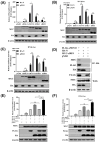
Fig 7. PRRSV N positively regulates IFN-β signaling pathway by interaction with NONO.
(A) PAM-KNU cells were cotransfected with 1 μg of pXJ41-sNONO and 1 μg of pXJ41-N or pXJ41 for 24 h and treated with 1 μg/mL poly (I:C) for 6 h. Cells were harvested and total RNA was extracted. The mRNA level of IFN-β was analyzed using real-time PCR (n = 3 independent experiment, *p < 0.05, **p < 0.01, bar indicates mean). (B-C) HEK-293T cells were cotransfected with 0.3 μg of pXJ41-sNONO and 0.3 μg of pXJ41-N or pXJ41, along with 0.3 μg of pIFN-β-Luc or pPRDI-III-Luc and 0.03 μg of pRL-TK luciferase reporter for 24 h, and then treated with 1 μg/mL poly (I:C) for 6 h. Cells were harvested and IFN-β promoter (B) or PRD I-III promoter (C) activity was analyzed by a dual-luciferase reporter assay (n = 3 independent experiment, *p < 0.05, **p < 0.01, bar indicates mean).
Fig 8
Fig 8. NONO promotes IRF3-induced IFN-β expression through binding to PRRSV N protein.
(A) PAM-KNU cells were transfected with 0.5 μg of pXJ41-N or pXJ41 for 24 h, along with 0.5 μg of pXJ41-sRIG-I, pXJ41-sMAVS, pXJ41-sTBK1, or pXJ41-sIRF3. Cells were harvested and total RNA was extracted. The mRNA level of IFN-β was analyzed using real-time PCR (n = 3 independent experiment, *p < 0.05, **p < 0.01, bar indicates mean). Whole-cell lysates were immunoblotted with anti-MYC or anti-HA antibody. The same blot was incubated with β-actin antibody as a protein loading control (n = 3 independent experiment, one representative experiment is shown). (B) PAM-KNU cells were transfected with 0.3 μg of pXJ41-N or pXJ41 together with 0.3 μg of pXJ41-sRIG-I, pXJ41-sMAVS, pXJ41-sTBK1, or pXJ41-sIRF3 with 0.3 μg of pIFN-β-Luc and 0.03 μg of pRL-TK luciferase reporter for 24 h. The activation of IFN-β promoter was detected using a dual-luciferase reporter assay (n = 3 independent experiment, *p < 0.05, **p < 0.01, bar indicates mean). Whole-cell lysates were immunoblotted with anti-MYC or anti-HA antibody. The same blot was incubated with β-actin antibody as a protein loading control (n = 3 independent experiment, one representative experiment is shown). (C) HEK-293T cells were transfected with 0.3 μg of pXJ41-N or pXJ41 together with 0.3 μg of pXJ41-hRIG-I, pXJ41-hMAVS, pXJ41-hTBK1, or pXJ41-hIRF3 with 0.3 μg of pIFN-β-Luc and 0.03 μg of pRL-TK luciferase reporter for 24 h. The IFN-β promoter activity was detected using a dual-luciferase reporter assay (n = 3 independent experiment, *p < 0.05, **p < 0.01, bar indicates mean). Whole-cell lysates were immunoblotted with anti-MYC or anti-HA antibody. The same blot was incubated with β-actin antibody as a protein loading control (n = 3 independent experiment, one representative experiment is shown). (D) HEK-293T cells were transfected with 2 μg of pXJ41-N, 2 μg of pXJ41-sNONO and 2 μg of pXJ41-sIRF3 for 24 h. Cells were harvested and subjected to co-IP with anti-HA antibody. Immunocomplexes were analyzed by Western blotting using anti-HA, anti-FLAG and anti-MYC antibodies. WCL was also subjected to Western blotting using anti-HA, anti-FLAG, anti-MYC, or anti-β-actin antibody (n = 3 independent experiment, one representative experiment is shown). (E) PAM-KNU cells were cotransfected with 0.5 μg of pXJ41-sNONO and 0.5 μg of pXJ41-N, pXJ41-N(NLS) or pXJ41 for 24 h, along with 0.5 μg of pXJ41-sIRF3. Cells were harvested and total RNA was extracted. The mRNA level of IFN-β was analyzed using real-time PCR (n = 3 independent experiment, **p < 0.01, "ns" stands for not statistically significant, bar indicates mean). Whole-cell lysates were immunoblotted with anti-MYC, anti-FLAG, or anti-HA antibody. The same blot was incubated with β-actin antibody as a protein loading control (n = 3 independent experiment, one representative experiment is shown). (F) HEK-293T cells were cotransfected with 0.3 μg of pXJ41-sNONO and 0.3 μg of pXJ41-N, pXJ41-N(NLS) or pXJ41 together with 0.3 μg of pXJ41-sIRF3 for 24 h, along with 0.3 μg of pIFN-β-Luc and 0.03 μg of pRL-TK luciferase reporter. Cells were harvested and IFN-β promoter activity was analyzed by a dual-luciferase reporter assay (n = 3 independent experiment, **p < 0.01, "ns" stands for not statistically significant, bar indicates mean). Whole-cell lysates were immunoblotted with anti-MYC, anti-FLAG, or anti-HA antibody. The same blot was incubated with β-actin antibody as a protein loading control (n = 3 independent experiment, one representative experiment is shown).
Fig 9
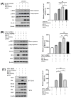
Fig 9. NONO enhances phosphorylation of IRF3 and interaction with IRF3 by binding to PRRSV N.
(A-B) HEK-293T cells were cotransfected with 1 μg of pXJ41-sNONO and pXJ41-N, pXJ41-N(NLS), or pXJ41 for 24 h, along with 1 μg/mL poly (I:C) treated for 6 h (A) or 1 μg of pXJ41-hTBK1 transfected (B). Cell lysates were analyzed by Western blotting using antibody against p-IRF3, IRF3, HA, FLAG, or β-actin (n = 3 independent experiment, one representative experiment is shown). The band intensities of p-IRF3 (long exposure) are shown as the relative protein expression levels, normalized with β-actin (n = 3 independent experiment, *p < 0.05, **p < 0.01, "ns" stands for not statistically significant, bar indicates mean). (C) HEK-293T cells were cotransfected with 2 μg of pXJ41-sNONO and 2 μg of pXJ41-N, pXJ41-N(NLS), or pXJ41 for 24 h, along with 2 μg of pXJ41-sIRF3. Cells were harvested and subjected to co-IP with anti-FLAG antibody. Immunocomplexes were analyzed by Western blotting using anti-MYC, anti-FLAG or anti-HA antibody. WCL was also subjected to Western blotting using anti-FLAG, anti-HA, anti-MYC, or anti-β-actin antibody (n = 3 independent experiment, one representative experiment is shown). The band intensities of IRF3 are shown as the relative protein expression levels, normalized with β-actin (n = 3 independent experiment, **p < 0.01, "ns" stands for not statistically significant, bar indicates mean).
Fig 10
Fig 10. The working model for NONO positively regulating IRF3-mediated IFN-β signaling pathway by detection of PRRSV N.

References

    1. Dokland T. The structural biology of PRRSV. Virus Res. 2010; 154(1–2):86–97. doi: 10.1016/j.virusres.2010.07.029 . - DOI - PMC - PubMed
    1. Schoggins JW, Rice CM. Interferon-stimulated genes and their antiviral effector functions. Curr Opin Virol. 2011; 1(6):519–525. doi: 10.1016/j.coviro.2011.10.008 . - DOI - PMC - PubMed
    1. Sun Q, Xu H, An T, Cai X, Tian Z, Zhang H. Recent Progress in Studies of Porcine Reproductive and Respiratory Syndrome Virus 1 in China. Viruses. 2023; 15(7). doi: 10.3390/v15071528 . - DOI - PMC - PubMed
    1. Zhang A, Duan H, Zhao H, Liao H, Du Y, Li L, et al.. Interferon-Induced Transmembrane Protein 3 Is a Virus-Associated Protein Which Suppresses Porcine Reproductive and Respiratory Syndrome Virus Replication by Blocking Viral Membrane Fusion. J Virol. 2020; 94(24). doi: 10.1128/JVI.01350-20 . - DOI - PMC - PubMed
    1. Liu XN, Li LW, Gao F, Jiang YF, Yuan WZ, Li GX, et al.. cGAS Restricts PRRSV Replication by Sensing the mtDNA to Increase the cGAMP Activity. Front Immunol. 2022; 13:887054. doi: 10.3389/fimmu.2022.887054 . - DOI - PMC - PubMed

MeSH terms